. 2022 Sep 21;8(10):990.
doi: 10.3390/jof8100990.

Caspofungin Affects Extracellular Vesicle Production and Cargo in Candida auris

Affiliations

Caspofungin Affects Extracellular Vesicle Production and Cargo in Candida auris

Rafaela F Amatuzzi et al. J Fungi (Basel). .

Abstract

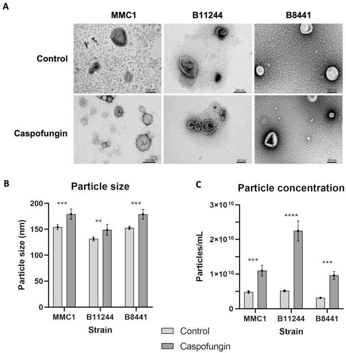
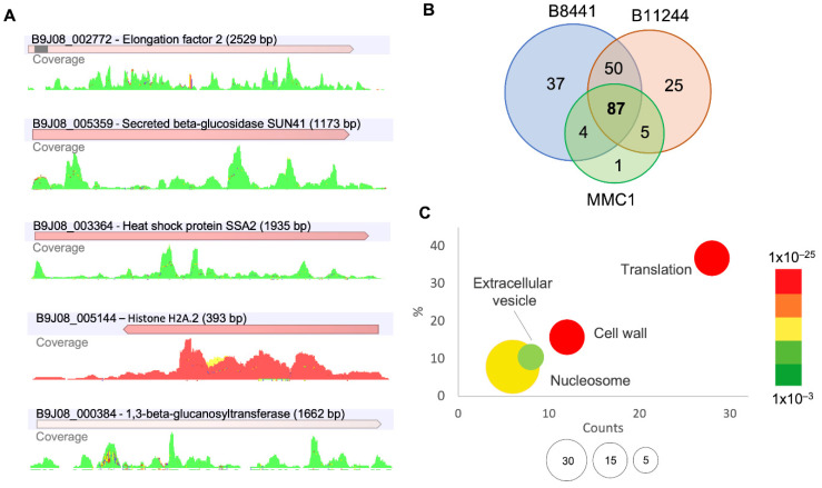
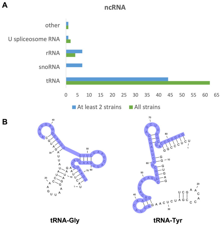
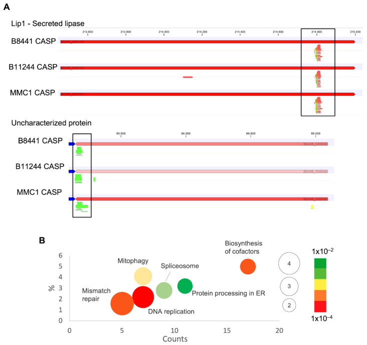
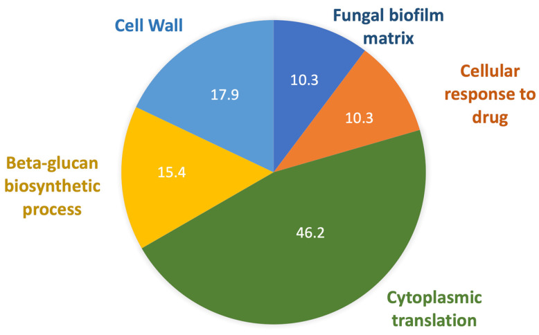
Antifungal resistance has become more frequent, either due to the emergence of naturally resistant species or the development of mechanisms that lead to resistance in previously susceptible species. Among these fungal species of global threat, Candida auris stands out for commonly being highly resistant to antifungal drugs, and some isolates are pan-resistant. The rate of mortality linked to C. auris infections varies from 28% to 78%. In this study, we characterized C. auris extracellular vesicles (EVs) in the presence of caspofungin, an echinocandin, which is the recommended first line antifungal for the treatment of infections due to this emerging pathogen. Furthermore, we also analyzed the protein and RNA content of EVs generated by C. auris cultivated with or without treatment with caspofungin. We observed that caspofungin led to the increased production of EVs, and treatment also altered the type and quantity of RNA molecules and proteins enclosed in the EVs. There were distinct classes of RNAs in the EVs with ncRNAs being the most identified molecules, and tRNA-fragments (tRFs) were abundant in each of the strains studied. We also identified anti-sense RNAs, varying from 21 to 55 nt in length. The differentially abundant mRNAs detected in EVs isolated from yeast subjected to caspofungin treatment were related to translation, nucleosome core and cell wall. The differentially regulated proteins identified in the EVs produced during caspofungin treatment were consistent with the results observed with the RNAs, with the enriched terms being related to translation and cell wall. Our study adds new information on how an echinocandin can affect the EV pathway, which is associated with the yeast cell being able to evade treatment and persist in the host. The ability of C. auris to efficiently alter the composition of EVs may represent a mechanism for the fungus to mitigate the effects of antifungal agents.

Keywords: Candida auris; RNA; drug resistance; extracellular vesicles; protein.


Conflict of interest statement

The authors declare that they have no conflict of interest.

Figures

Figure 1
Figure 1
Characterization of Candida auris extracellular vesicles (EVs). (A) Transmission electron microscopy images of EVs derived from MMC1, B11244 and B8441 strains under control conditions and caspofungin treatment. bar = 200 nm. (B) Particle size distribution bar chart for MMC1, B11244 and B8441 EVs from control (light grey) and caspofungin treatment (dark grey). The average EV size (y-axis) is indicated in nm. The x-axis indicates the strains studied, where *** p < 0.005 and ** p < 0.05, by individual t-tests followed by Bonferroni’s multicomparisons test for three independent experiments. (C) A particle concentration bar chart related to EVs concentration calculated the three strains from control and caspofungin treatment; the y-axis indicates the particles concentration, *** p < 0.001 and **** p < 0.0001, by one-way Anova followed by Bonferroni’s multicomparisons test for three independent experiments. The EV size is indicated in nm.
Figure 2
Figure 2
mRNAs present in C. auris EVs. (A) Reads coverage profile of transcripts enriched in EVs isolated after caspofungin treatment; forward reads (green), reverse reads (red) and non-specific match (yellow). (B) Venn chart of the mRNAs present in the EVs from the strains used in this study. (C) Gene ontology bubble chart, with the common transcripts identified in the EVs from the three strains, with the terms enriched in the presence of caspofungin. X-axis, the number of counts for the terms identified, Y-axis, the percentage of terms in the analysis; the bubble size reflects the fold-enrichment of the term after Fisher-exact test was applied and the color code reflect the p-value of the terms.
Figure 3
Figure 3
ncRNAs present in C. auris EVs. (A) Bar chart describing the most prevalent classes of ncRNAs identified in the EVs in the B8441, B11244 and MMC1 strains in green and the ncRNA identified in at least two strains in blue. (B) tRNA structure and sequence for the tRNA-Gly and tRNA-Tyr. Light purple highlights the region sequenced in our analysis, demonstrating that they are tRNA-derived fragments (tfRNAs).
Figure 4
Figure 4
anti-sense RNAs present in C. auris EVs. (A) Specific regions of reads aligning in transcripts from EVs isolated after caspofungin treatment, in red reverse reads, and in green forward reads. (B) Gene ontology bubble chart of the targets of the asRNAs identified in the EVs from at least two of the studied strains, with the terms enriched in the presence of caspofungin. X-axis, the number of counts for the terms identified; Y-axis is the percentage of terms in the analysis, and the bubble size reflects the fold-enrichment of the term after a Fisher’s-exact test was applied and the color code reflects the p-value of the terms.
Figure 5
Figure 5
Proteins present in C. auris EVs. Pie chart of gene ontology terms identified in the EVs from all the strains enriched when the cells were treated with caspofungin.

Similar articles

Cited by

References

    1. Satoh K., Makimura K., Hasumi Y., Nishiyama Y., Uchida K., Yamaguchi H. Candida Auris Sp. Nov., a Novel Ascomycetous Yeast Isolated from the External Ear Canal of an Inpatient in a Japanese Hospital. Microbiol. Immunol. 2009;53:41–44. doi: 10.1111/j.1348-0421.2008.00083.x. - DOI - PubMed
    1. Pandya N., Cag Y., Pandak N., Pekok A.U., Poojary A., Ayoade F., Fasciana T., Giammanco A., Caskurlu H., Rajani D.P., et al. International Multicentre Study of Candida Auris Infections. J. Fungi. 2021;7:878. doi: 10.3390/jof7100878. - DOI - PMC - PubMed
    1. Garcia-Bustos V., Salavert M., Ruiz-Gaitán A.C., Cabañero-Navalon M.D., Sigona-Giangreco I.A., Pemán J. A Clinical Predictive Model of Candidaemia by Candida Auris in Previously Colonized Critically Ill Patients. Clin. Microbiol. Infect. 2020;26:1507–1513. doi: 10.1016/j.cmi.2020.02.001. - DOI - PubMed
    1. Chowdhary A., Sharma C., Meis J.F. Candida Auris: A Rapidly Emerging Cause of Hospital-Acquired Multidrug-Resistant Fungal Infections Globally. PLoS Pathog. 2017;13:e1006290. doi: 10.1371/journal.ppat.1006290. - DOI - PMC - PubMed
    1. De Jong A.W., Hagen F. Attack, Defend and Persist: How the Fungal Pathogen Candida Auris Was Able to Emerge Globally in Healthcare Environments. Volume 184 Springer; Heidelberg, The Netherlands: 2019. - PubMed