. 2022 Sep 22;8(10):997.
doi: 10.3390/jof8100997.

The Small Ras Superfamily GTPase Rho4 of the Maize Anthracnose Fungus Colletotrichum graminicola Is Required for β-1,3-glucan Synthesis, Cell Wall Integrity, and Full Virulence

Affiliations

The Small Ras Superfamily GTPase Rho4 of the Maize Anthracnose Fungus Colletotrichum graminicola Is Required for β-1,3-glucan Synthesis, Cell Wall Integrity, and Full Virulence

Ely Oliveira-Garcia et al. J Fungi (Basel). .

Abstract

Small Ras superfamily GTPases are highly conserved regulatory factors of fungal cell wall biosynthesis and morphogenesis. Previous experiments have shown that the Rho4-like protein of the maize anthracnose fungus Colletotrichum graminicola, formerly erroneously annotated as a Rho1 protein, physically interacts with the β-1,3-glucan synthase Gls1 (Lange et al., 2014; Curr. Genet. 60:343-350). Here, we show that Rho4 is required for β-1,3-glucan synthesis. Accordingly, Δrho4 strains formed distorted vegetative hyphae with swellings, and exhibited strongly reduced rates of hyphal growth and defects in asexual sporulation. Moreover, on host cuticles, conidia of Δrho4 strains formed long hyphae with hyphopodia, rather than short germ tubes with appressoria. Hyphopodia of Δrho4 strains exhibited penetration defects and often germinated laterally, indicative of cell wall weaknesses. In planta differentiated infection hyphae of Δrho4 strains were fringy, and anthracnose disease symptoms caused by these strains on intact and wounded maize leaf segments were significantly weaker than those caused by the WT strain. A retarded disease symptom development was confirmed by qPCR analyses. Collectively, we identified the Ras GTPase Rho4 as a new virulence factor of C. graminicola.

Keywords: Colletotrichum graminicola; Rho GTPases; Zea mays; appressoria; fungal cell walls; hyphopodia; penetration defect; virulence factor; β-1,3-glucan synthesis.


Conflict of interest statement

The authors declare no conflict of interest.

Figures

Figure 1
Figure 1
Phylogenetic tree and organization of conserved domains in Rho proteins of C. graminicola, as compared with Rho GTPase proteins of yeasts and other filamentous fungi. (A) Size and positions of G-boxes and prenylation sites of Rho proteins of C. graminicola and the filamentous and yeast model fungi A. niger and S. cerevisiae. Amino acid (aa) numbers of the Rho proteins are given on the right side. (B) Sequence logo indicating amino acid sequence conservation of G-boxes and prenylation sites. Comparisons were made using the amino acid sequences of C. graminicola, of the model filamentous ascomycetes Magnaporthe oryzae, N. crassa, and Aspergillus niger, and of the yeasts S. cerevisiae and S. pombe. (C) Rooted phylogenetic tree indicates close relatedness of Rho1–Rho4 proteins of different filamentous ascomycetes and yeasts. The S. cerevisiae Ran1 protein served as an outgroup. Solid and open circles indicate bootstraps higher or lower than 80.
Figure 2
Figure 2
Relevance of C. graminicola Rho4 for growth, hyphal morphology, and sensitivity to Caspofungin. (A) Growth of the WT, Δrho4, and ectopic strains on OMA or OMA supplemented with osmolytes (OMA + KCl or OMA + sorbitol). Arrowheads point at salmon-colored acervuli with conidia. Arrows indicate the hyper-pigmented Δrho4 strain. Plates were photographed at 4 dpi. (B) Growth of WT, ectopic, and Δrho4 strains on OMA. Error bars indicate SDs. (C) Microscopy indicated severe cell wall defects of Δrho4 strains. As compared with the WT strain, DIC microscopy of the Δrho4 strain displayed massive ballooning of vegetative hyphae (Δrho4 DIC, arrows; compare with WT DIC). Bright-field microscopy showed intrahyphal hyphae (Δrho4 BF, asterisk) developing within hyphae (Δrho4 BF, arrowhead). While Calcofluor White staining of WT hyphae revealed clear septation (WT CFW, arrowheads), brightly stained swellings (Δrho4 CFW asterisk) and diffuse septa (Δrho4 CFW arrowheads) were seen in hyphae of the mutant. Microscopy was done at 4 dpi. Scale bars are 10 μm. (D) Colony growth of WT and Δrho4 strains on PDA + KCl, and on PDA + KCl amended with the antifungals Caspofungin or Nystatin. Growth of the WT strain is strongly retarded by all antifungals. The Δrho4 strain is not inhibited by Caspofungin, but growth is completely blocked by Nystatin. The size of the agar block used as a Δrho4 inoculum is clearly visible. Plates were photographed at 7 dpi. (E) Growth inhibition ratio of Δrho4 vs. WT strain. Growth on media containing antifungals is expressed relative to growth on PDA + KCl. Error bars indicate SDs.
Figure 3
Figure 3
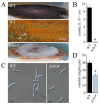
Asexual sporulation of the C. graminicola WT and Δrho4 strains on PDA amended with KCl. (A) After 7 dpi the WT had formed a dark mycelium, which developed orange-colored acervuli (arrows) by 14 dpi. The Δrho4 strain developed aerial mycelium without visible acervuli. (B) Quantification of conidia formed by the WT and Δrho4 strains. (C) Shape of conidia formed by the WT and Δrho4 strains. Scale bars are 20 μm. (D) Length of conidia formed by the WT and Δrho4 strains. Error bars in B and D are SDs. Asterisks indicate statistically significant differences (p ≤ 0.05).
Figure 4
Figure 4
Chitin and β-1,3-glucan abundance in vegetative hyphae of the WT and Δrho4 mutant. (A) WT strain and Δrho4 hyphae were stained with Aniline Blue fluorochrome and Alexa Fluor 647-Wheat Germ Agglutinin conjugate to detect β-1,3-glucan and chitin, respectively. While hyphae of the WT strain showed strong β-1,3-glucan labeling (arrowheads), those of Δrho4 strains did not. By contrast, chitin was hardly observed in the WT strain, but abundant in Δrho4 hyphae, with prominent labeling at hyphal protrusions (arrows). Scale bars are 10 μm. (B) Quantification of fluorescence intensity in hyphae labelled as in (A), from the hyphal tip to subapical regions. Virtual line scans confirm strong β-1,3-glucan labeling of the WT and strong chitin labeling of Δrho4 hyphae. (C) The β-1,3-glucan-to-chitin fluorescence ratios of the WT and Δrho4 strains show that the proportion of β-glucan is substantially reduced in Δrho4 strains. Error bars indicate SDs.
Figure 5
Figure 5
Invasive growth of the WT and strains with homologous and ectopic integration of the deletion cassette in race tubes. (A) Race tubes filled with PDA osmotically stabilized with KCl visualized invasive growth of the WT and Δrho4 strains. (B) Quantification of invasive growth as dependent of the agar concentration. Error bars are SDs.
Figure 6
Figure 6
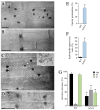
Δrho4 strains are defective in infection structure differentiation on maize leaves. (AD) DIC microscopy of the infection process of C. graminicola WT and Δrho4 strains. Conidia (A,C) of the WT strain germinate and differentiate melanized appressoria ((A), app), which invade the epidermal host cell and differentiate infection hyphae ((B), ih). The Δrho4 mutant, by contrast, exhibits bipolar germination ((C), arrows), which form long ((C), arrowheads) and often coiled hyphae ((C), dashed box) before they differentiate hyphopodia ((C), hy). Several hyphopodia of the Δrho4 mutant germinate laterally ((C), insert; asterisks and numbers 1–3 indicate first–third hyphopodium; arrowheads with numbers 1 and 2 indicate first and second lateral germ tube). Not only hyphopodia, but also appressoria, are formed ((C), app). After penetration, the Δrho4 mutant forms fringy infection hyphae with numerous small branches ((D), ih). Scale bars in (AC) are 20 μm; scale bar in (D) is 50 μm. (E) Rate of bipolar germination of WT and Δrho4 conidia. Error bars are SDs; asterisk indicates statistically significant differences (t-test, p ≤ 0.05). (F) Ratio of hyphopodia to appressoria formed by WT and Δrho4 strains. Error bars are SDs; asterisk indicates statistically significant differences (t-test, p ≤ 0.05). (G) Appressorium, hyphopodium, and infection hypha differentiation by WT and Δrho4 strains. Error bars are SDs; asterisks indicate statistically significant differences of corresponding structures (t-test, p ≤ 0.05).
Figure 7
Figure 7
Δrho4 strains are nonpathogenic on wounded and non-wounded leaves. (A) Disease symptoms on non-wounded and wounded maize leaves after inoculation with the WT and Δrho4 strains. Mock-inoculated leaves were treated with 0.01% (v/v) Tween 20 and served as controls (mock). Photographs were taken at 4 dpi. (B) Quantification of fungal development on non-wounded and wounded maize leaves by qPCR. Three independent biological and four technical replicates were analyzed for WT and Δrho4 strains. Error bars are SDs. Asterisks indicate statistically significant differences (t-test, p ≤ 0.05).

Similar articles

References

    1. Cosio E.G., Feger M., Miller C.J., Antelo L., Ebel J. High affinity binding of fungal β-glucan elicitors to cell membranes of species of the plant family Fabaceae. Planta. 1996;200:92–99. doi: 10.1007/BF00196654. - DOI
    1. Mithöfer A., Fliegmann J., Neuhaus-Uri G., Schwarz H., Ebel J. The hepta-β-glucoside elicitor-binding proteins from legumes represent a putative receptor family. Biol. Chem. 2000;381:705–713. doi: 10.1515/BC.2000.091. - DOI - PubMed
    1. Böhm H., Albert I., Fan L., Reinhard A., Nürnberger T. Immune receptor complexes at the plant cell surface. Curr. Opin. Plant Biol. 2014;20:47–54. doi: 10.1016/j.pbi.2014.04.007. - DOI - PubMed
    1. Fesel P.H., Zuccaro A. β-glucan: Crucial component of the fungal cell wall and elusive MAMP in plants. Fungal Genet. Biol. 2016;90:53–60. doi: 10.1016/j.fgb.2015.12.004. - DOI - PubMed
    1. Oliveira-Garcia E., Deising H.B. Infection structure-specific expression of β-1,3-glucan synthase is essential for pathogenicity of Colletotrichum graminicola and evasion of β-glucan-triggered immunity. Plant Cell. 2013;25:2356–2378. doi: 10.1105/tpc.112.103499. - DOI - PMC - PubMed

LinkOut - more resources