. 2022 Sep 29;8(10):1032.
doi: 10.3390/jof8101032.

The Transcription Factor CsAtf1 Negatively Regulates the Cytochrome P450 Gene CsCyp51G1 to Increase Fludioxonil Sensitivity in Colletotrichum siamense

Affiliations

The Transcription Factor CsAtf1 Negatively Regulates the Cytochrome P450 Gene CsCyp51G1 to Increase Fludioxonil Sensitivity in Colletotrichum siamense

Xiaoling Guan et al. J Fungi (Basel). .

Abstract

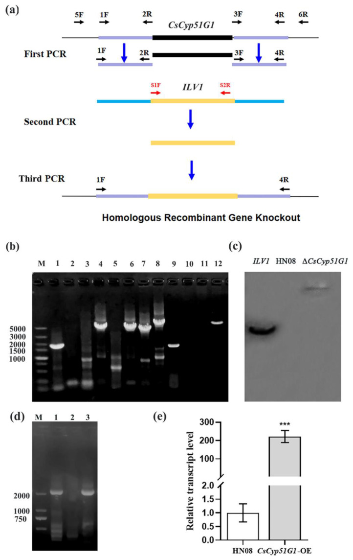
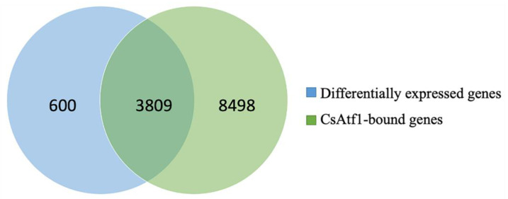
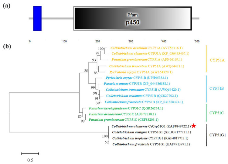
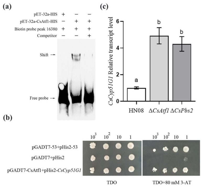
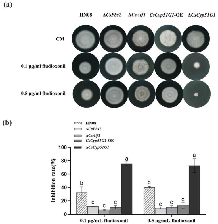
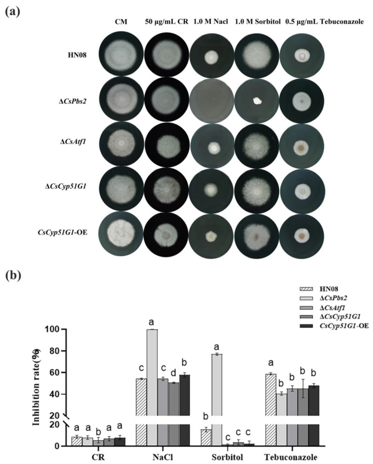
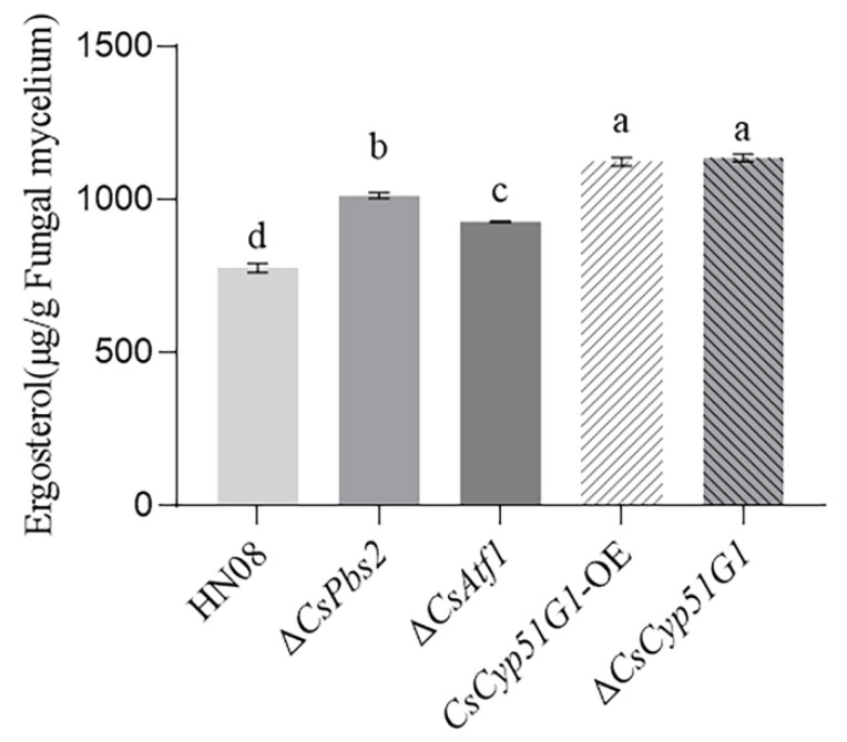
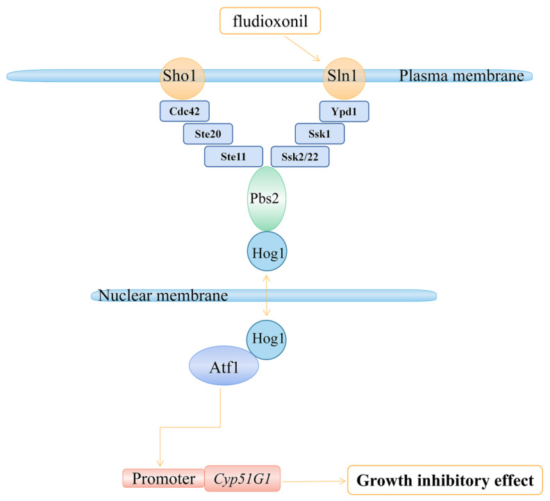
Previous studies have shown that the high-osmolarity glycerol mitogen-activated protein kinase (HOG MAPK) signaling pathway and its downstream transcription factor CsAtf1 are involved in the regulation of fludioxonil sensitivity in C. siamense. However, the downstream target genes of CsAtf1 related to the fludioxonil stress response remain unclear. Here, we performed chromatin immunoprecipitation sequencing (ChIP-Seq) and high-throughput RNA-sequencing (RNA-Seq) to identify genome-wide potential CsAtf1 target genes. A total of 3809 significantly differentially expressed genes were predicted to be directly regulated by CsAtf1, including 24 cytochrome oxidase-related genes. Among them, a cytochrome P450-encoding gene, designated CsCyp51G1, was confirmed to be a target gene, and its transcriptional expression was negatively regulated by CsAtf1, as determined using an electrophoretic mobility shift assay (EMSA), a yeast one-hybrid (Y1H) assay, and quantitative real-time PCR (qRT-PCR). Moreover, the overexpression mutant CsCYP51G1 of C. siamense exhibited increased fludioxonil tolerance, and the CsCYP51G1 deletion mutant exhibited decreased fludioxonil resistance, which revealed that CsCyp51G1 is involved in fludioxonil sensitivity regulation in C. siamense. However, the cellular ergosterol content of the mutants was not consistent with the phenotype of fludioxonil sensitivity, which indicated that CsCyp51G1 regulates fludioxonil sensitivity by affecting factors other than the ergosterol level in C. siamense. In conclusion, our data indicate that the transcription factor CsAtf1 negatively regulates the cytochrome P450 gene CsCyp51G1 to increase fludioxonil sensitivity in C. siamense.

Keywords: ChIP-Seq; Colletotrichum siamense; CsAtf1; CsCyp51G1; RNA-Seq; cytochrome P450; fludioxonil sensitivity.


Conflict of interest statement

The authors declare no conflict of interest.

Figures

Figure 1
Figure 1
Verification of the gene deletion mutant ΔCsCyp51G1 and overexpression strain CsCyp51G1-OE. (a) Schematic diagram of CsCyp51G1 gene deletion and primers used to verify the gene replacement event. (b) Confirmation of the ΔCsCyp51G1 mutant and CsCyp51G1-OE strain by PCR amplification. M: DNA DL 5000 marker; A 1712 bp fragment of the CsCyp51G1 gene coding sequence was amplified by the primer pair CsCyp51G1-OF/OR in both HN08 (lane 1) and CsCyp51G1-OE (lane 9) but not ΔCsCyp51G1 (lane 5). A 4217-bp fragment containing the upstream sequence of the CsCyp51G1 gene and a partial ILV1 gene sequence was amplified by the primer pair 5F/S2R from ΔCsCyp51G1 (lane 6) but not HN08 (lane 2) or CsCyp51G1-OE (lane 10). A 4018-bp fragment containing the downstream sequence of the CsCyp51G1 gene and a partial ILV1 gene sequence was amplified by the primer pair S1F/6R from ΔCsCyp51G1 (lane 7) but not HN08 (lane 3) or CsCyp51G1-OE (lane 11). A 5267-bp fragment from ΔCsCyp51G1 (lane 8) and a 4313-bp fragment from HN08 (lane 4) and CsCyp51G1-OE (lane 12) were amplified by the primer pair 5F/6R, which confirmed that the 1712-bp fragment of the CsCyp51G1 gene was replaced by the 2817-bp fragment of the ILV1 gene. (c) Southern blot analysis of total genomic DNA samples from WT and ΔCsCyp51G1 were digested with EcoRI. The probe was an ILV1 PCR fragment amplified from the sulfonylurea resistance cassette of the plasmid pCX62-S. (d) PCR with the primer pair RP27F/CsCyp51G1-OR amplified a 2189-bp fragment from the plasmid pXY203-RP27-Cyp51G1 and the CsCyp51G1-OE overexpression strain but not the wild-type strain. (e) Relative expression of CsCyp51G1 determined by qRT-PCR in CsCyp51G1-OE strains and WT HN08. Expression levels were normalized using ACT expression levels as controls. Data were collected from three technical replicates. Error bars represent the standard deviations, *** indicated significant differences at p < 0.001 (Student’s paired two-tailed t-test).-b.
Figure 2
Figure 2
Venn diagram of significantly differentially expressed genes in the ΔCsAtf1 mutant and genes bound directly by the transcription factor CsAtf1.
Figure 3
Figure 3
Protein domains and phylogenetic analysis of CsCyp51G1. (a) SMART analysis of the CsCyp51G1 protein domain. (b) Phylogenetic analysis of CsCyp51G1 and its homologues from other fungal species. The phylogenetic tree was constructed with MEGA 6.0 using the maximum likelihood method. The CsCyp51G1 protein amino acid sequence in this study is indicated with a red star.
Figure 4
Figure 4
Analysis of the binding and expression relationship between CsAtf1 and CsCyp51G1. (a) Detection of the binding relationship between the CsAtf1 protein and the promoter regions of CsCyp51G1 by EMSA. +, protein added, −, protein not added. (b) Y1H Gold confirmed that the transcription factor CsAtf1 binds to the promoter of CsCyp51G1. The representative growth status of different cell culture dilutions (1:1, 1:10, 1:100, 1:1000) on SD/-Leu/-Trp/-His agar medium without and with 80 mM 3-AT is shown. (c) Relative expression analysis of CsCyp51G1 in the wild-type HN08, ΔCsPbs2, and ΔCsAtf1 strains. Different letters (a, b) represent significant difference at p < 0.01 (One-way ANOVA and Duncan’s test).
Figure 5
Figure 5
Comparison of fludioxonil resistance among the WT strain HN08, ΔCsPbs2, ΔCsAtf1, CsCyp51G1-OE, and ΔCsCyp51G1. (a) Mycelial growth of the tested strains cultured in CM containing different concentrations of fludioxonil for 5 d. (b) Growth inhibition rate of the tested strains under different concentrations of fludioxonil. The growth inhibition rate is relative to the growth rate of each untreated control [(diameter of untreated strain − diameter of treated strain)/(diameter of untreated strain × 100%)]. Three repeats were performed. The error bars show the SD value, and different letters indicate significant difference at p < 0.01 (One-way ANOVA and Duncan’s test).
Figure 6
Figure 6
Comparison of responses to various stresses among HN08, ΔCsPbs2, ΔCsAtf1, CsCyp51G1-OE, and ΔCsCyp51G1. (a) Mycelial growth of HN08, ΔCsPbs2, ΔCsAtf1, CsCyp51G1-OE, and ΔCsCyp51G1 on CM containing 1 M NaCl, 1 M sorbitol, 50 μg/mL Congo red, and 0.5 μg/mL tebuconazole for 5 d. Approximately 1 × 10⁶ conidia of each strain were inoculated onto the center of the CM plates and grown for 5 d. (b) Growth inhibition rates of the tested strains under different stresses. The growth inhibition rate is relative to the growth rate of each untreated control [(diameter of untreated strain − diameter of treated strain)/(diameter of untreated strain × 100%)]. Three repeats were performed. Error bars represent the standard deviations. Different letters (a, b, c, d) represent significant difference at p < 0.01 (One-way ANOVA and Duncan’s test).
Figure 7
Figure 7
Ergosterol content in HN08, ΔCsPbs2, ΔCsAtf1, CsCyp51G1-OE, and ΔCsCyp51G1. The error bars show the SD values, and different letters (a, b, c, d) represent significant difference at p < 0.01 (One-way ANOVA and Duncan’s test).
Figure 8
Figure 8
Proposed model from our study showing that CsAtf1 downregulates CsCyp51G1 expression levels, leading to fludioxonil sensitivity.

Similar articles

Cited by

References

    1. O’Connell R.J., Thon M.R., Hacquard S., Amyotte S.G., Kleemann J., Torres M., Damm U., Buiate E.A., Epstein L., Alkan N., et al. Lifestyle transitions in plant pathogenic Colletotrichum fungi deciphered by genome and transcriptome analyses. Nat. Genet. 2012;44:1060–1065. doi: 10.1038/ng.2372. - DOI - PMC - PubMed
    1. Dean R., Van Kan J.A., Pretorius Z.A., Hammond-Kosack K.E., Di Pietro A., Spanu P.D., Rudd J.J., Dickman M., Kahmann R., Ellis J., et al. The top 10 fungal pathogens in molecular plant pathology. Mol. Plant Pathol. 2012;13:414–430. doi: 10.1111/j.1364-3703.2011.00783.x. - DOI - PMC - PubMed
    1. Cao X., Xu X., Che H., West J.S., Luo D. Eight Colletotrichum species, including a novel species, are associated with areca palm anthracnose in Hainan, China. Plant Dis. 2020;104:1369–1377. doi: 10.1094/PDIS-10-19-2077-RE. - DOI - PubMed
    1. Qin L.P., Zhang Y., Su Q., Chen Y.L., Nong Q., Xie L., Yu G.M., Huang S.L. First report of anthracnose of Mangifera indica caused by Colletotrichum scovillei in China. Plant Dis. 2019;103:1043. doi: 10.1094/PDIS-11-18-1980-PDN. - DOI
    1. Huang R., Sun W., Wang L., Li Q., Huang S., Tang L., Guo T., Mo J., Hsiang T. Identification and characterization of Colletotrichum species associated with anthracnose disease of banana. Plant Pathol. 2021;70:1827–1837. doi: 10.1111/ppa.13426. - DOI

LinkOut - more resources