

Sonographic Assessment of Uterine Biometry for the Diagnosis of Diffuse Adenomyosis in a Tertiary Outpatient Clinic
- PMID: 36294711
- PMCID: PMC9604640
- DOI: 10.3390/jpm12101572
Sonographic Assessment of Uterine Biometry for the Diagnosis of Diffuse Adenomyosis in a Tertiary Outpatient Clinic
Abstract
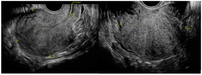
Background: to compare several uterine biometric parameters at transvaginal ultrasound (TVUS) between adenomyosis and non-adenomyosis uteri and evaluate their role for the diagnosis of diffuse adenomyosis.
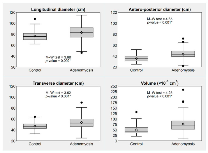
Methods: prospective observational study conducted between the 1 February 2022 and the 30 April 2022. In this case, 56 patients with TVUS diagnosis of adenomyosis were included. A 1:1 ratio age and parity-matched group of non-adenomyosis patients was selected. We compared sonographic uterine biometric parameters (longitudinal (LD), anteroposterior (APD) and transverse (TD) diameters, volume, simple and complex diameter ratios) and investigated their diagnostic performance.
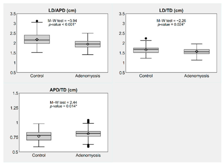
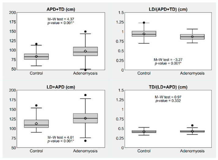
Results: all sonographic parameters were significantly different between the study groups, except for TD/(LD+APD). Optimal cut-off values of APD and LD/APD showed the best sensitivity and specificity. APD diameter equal or superior to 39.5 mm (95% CI, 36.2-42.8) had sensitivity of 0.70 (95% CI, 0.57-0.80), specificity of 0.71 (95% CI, 0.59-0.82) and accuracy of 0.75 (95% CI, 0.66-0.84). LD/APD equal or inferior to 2.05 (95% CI, 1.96-2.13) showed sensitivity and specificity of 0.70 (95% CI, 0.57-0.80) each and accuracy of 0.72 (95% CI, 0.62-0.81).
Conclusions: several biometric uterine parameters at TVUS in fertile-aged women were statistically different between adenomyosis and non-adenomyosis uteri, though their optimal cut-off values showed low accuracy in diagnosing adenomyosis.
Keywords: adenomyosis; biometry; diagnosis; globular uterus; ultrasonography; ultrasound.
Conflict of interest statement
The authors declare no conflict of interest.
Figures
References
-
- van den Bosch T., Dueholm M., Leone F.P.G., Valentin L., Rasmussen C.K., Votino A., Van Schoubroeck D., Landolfo C., Installé A.J., Guerriero S., et al. Terms, definitions and measurements to describe sonographic features of myometrium and uterine masses: A consensus opinion from the Morphological Uterus Sonographic Assessment (MUSA) group. Ultrasound Obstet. Gynecol. 2015;46:284–298. doi: 10.1002/uog.14806. - DOI - PubMed
-
- Exacoustos C., Morosetti G., Conway F., Camilli S., Martire F.G., Lazzeri L., Piccione E., Zupi E. New Sonographic Clas-sification of Adenomyosis: Do Type and Degree of Adenomyosis Correlate to Severity of Symptoms? J. Minim. Invasive Gynecol. 2020;27:1308–1315. doi: 10.1016/j.jmig.2019.09.788. - DOI - PubMed
LinkOut - more resources
Full Text Sources
Research Materials
Miscellaneous
