

Quantifying Intracellular Viral Pathogen: Specimen Preparation, Visualization and Quantification of Multiple Immunofluorescent Signals in Fixed Human Airway Epithelium Cultured at Air-Liquid Interface
- PMID: 36294807
- PMCID: PMC9605096
- DOI: 10.3390/jpm12101668
Quantifying Intracellular Viral Pathogen: Specimen Preparation, Visualization and Quantification of Multiple Immunofluorescent Signals in Fixed Human Airway Epithelium Cultured at Air-Liquid Interface
Abstract
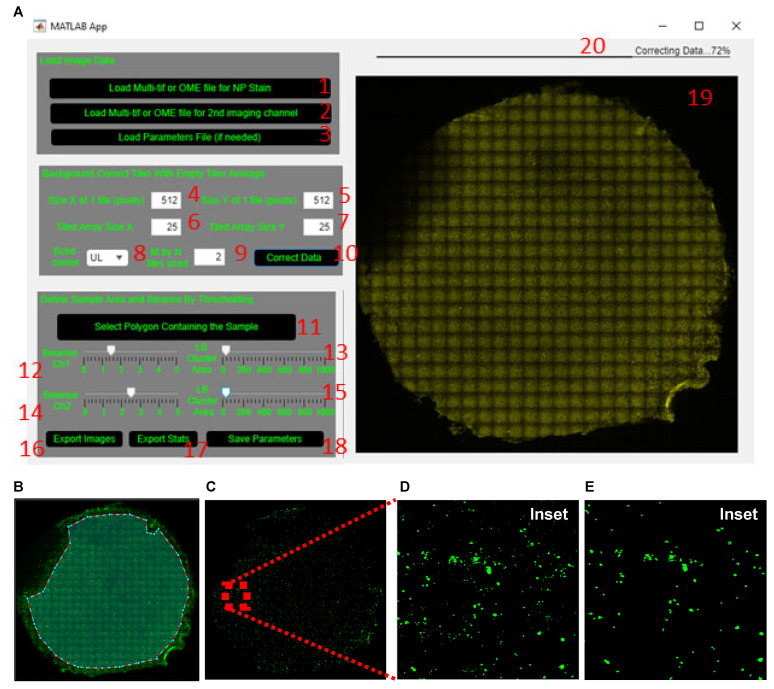
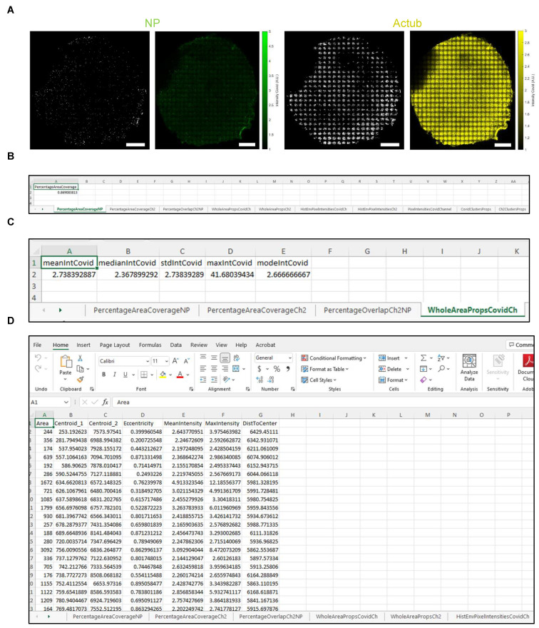
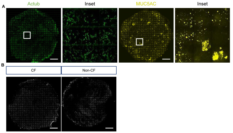
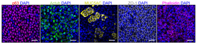
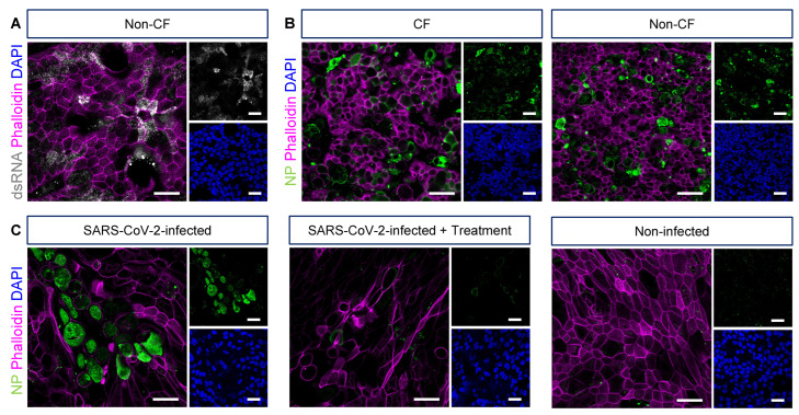
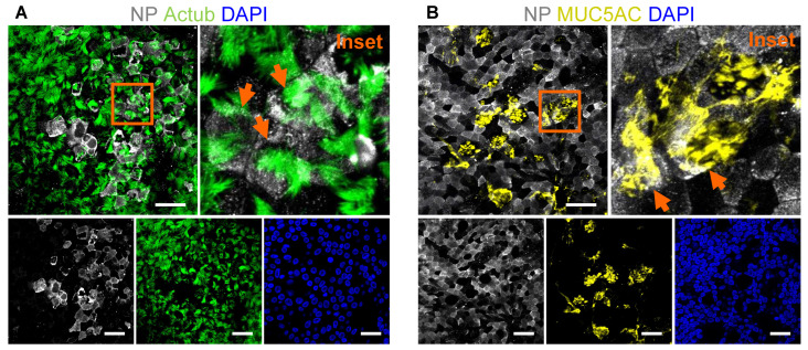
Infection control and aggressive antibiotic therapy play an important role in the management of airway infections in individuals with cystic fibrosis (CF). The responses of airway epithelial cells to pathogens are likely to contribute to the pathobiology of CF lung disease. Primary airway epithelial cells obtained from individuals with CF, cultured and differentiated at air-liquid interface (ALI), effectively mimic the structure and function of the in vivo airway epithelium. With the recent respiratory viral pandemics, ALI cultures were extensively used to model respiratory infections in vitro to facilitate physiologically relevant respiratory research. Immunofluorescence staining and imaging were used as an effective tool to provide a fundamental understanding of host-pathogen interactions and for exploring the therapeutic potential of novel or repurposed drugs. Therefore, we described an optimized quantitative fluorescence microscopy assay for the wholemount staining and imaging of epithelial cell markers to identify distinct cell populations and pathogen-specific targets in ALI cultures of human airway epithelial cells grown on permeable support insert membranes. We present a detailed methodology using a graphical user interface (GUI) package to quantify the detected signals on a tiled whole membrane. Our method provided an imaging strategy of the entire membrane, overcoming the common issue of undersampling and enabling unbiased quantitative analysis.
Keywords: SARS-CoV-2; air-liquid interface; airway infection; cystic fibrosis; quantification; tiled imaging; wholemount immunofluorescence.
Conflict of interest statement
The authors declare no conflict of interest.
Figures
Similar articles
-
Characterization of pediatric cystic fibrosis airway epithelial cell cultures at the air-liquid interface obtained by non-invasive nasal cytology brush sampling.Respir Res. 2017 Dec 28;18(1):215. doi: 10.1186/s12931-017-0706-7. Respir Res. 2017. PMID: 29282053 Free PMC article.
-
Immunofluorescence-Mediated Detection of Respiratory Virus Infections in Human Airway Epithelial Cultures.Curr Protoc. 2022 Jun;2(6):e453. doi: 10.1002/cpz1.453. Curr Protoc. 2022. PMID: 35671174 Free PMC article.
-
A toolbox for studying respiratory viral infections using air-liquid interface cultures of human airway epithelial cells.Am J Physiol Lung Cell Mol Physiol. 2021 Jul 1;321(1):L263-L280. doi: 10.1152/ajplung.00141.2021. Epub 2021 May 19. Am J Physiol Lung Cell Mol Physiol. 2021. PMID: 34010062
-
Elucidating the Interaction of CF Airway Epithelial Cells and Rhinovirus: Using the Host-Pathogen Relationship to Identify Future Therapeutic Strategies.Front Pharmacol. 2018 Nov 7;9:1270. doi: 10.3389/fphar.2018.01270. eCollection 2018. Front Pharmacol. 2018. PMID: 30464745 Free PMC article. Review.
-
Invited review: human air-liquid-interface organotypic airway tissue models derived from primary tracheobronchial epithelial cells-overview and perspectives.In Vitro Cell Dev Biol Anim. 2021 Feb;57(2):104-132. doi: 10.1007/s11626-020-00517-7. Epub 2020 Nov 11. In Vitro Cell Dev Biol Anim. 2021. PMID: 33175307 Free PMC article. Review.
Cited by
-
Molecular and Functional Characteristics of Airway Epithelium under Chronic Hypoxia.Int J Mol Sci. 2023 Mar 30;24(7):6475. doi: 10.3390/ijms24076475. Int J Mol Sci. 2023. PMID: 37047450 Free PMC article.
-
Q1291H-CFTR molecular dynamics simulations and ex vivo theratyping in nasal epithelial models and clinical response to elexacaftor/tezacaftor/ivacaftor in a Q1291H/F508del patient.Front Mol Biosci. 2023 Jun 1;10:1148501. doi: 10.3389/fmolb.2023.1148501. eCollection 2023. Front Mol Biosci. 2023. PMID: 37325471 Free PMC article.
References
-
- Dijkman R., Jebbink M.F., Koekkoek S.M., Deijs M., Jónsdóttir H.R., Molenkamp R., Ieven M., Goossens H., Thiel V., van der Hoek L. Isolation and Characterization of Current Human Coronavirus Strains in Primary Human Epithelial Cell Cultures Reveal Differences in Target Cell Tropism. J. Virol. 2013;87:6081–6090. doi: 10.1128/JVI.03368-12. - DOI - PMC - PubMed
-
- Tran B.M., Grimley S.L., McAuley J.L., Hachani A., Earnest L., Wong S.L., Caly L., Druce J., Purcell D.F., Jackson D.C., et al. Air-Liquid-Interface Differentiated Human Nose Epithelium: A Robust Primary Tissue Culture Model of SARS-CoV-2 Infection. Int. J. Mol. Sci. 2022;23:835. doi: 10.3390/ijms23020835. - DOI - PMC - PubMed
Grants and funding
LinkOut - more resources
Full Text Sources
Miscellaneous
