

Review
doi: 10.3390/jpm12101733.
Clinical Application of Ultra-High-Frequency Ultrasound
Affiliations
- PMID: 36294872
- PMCID: PMC9605054
- DOI: 10.3390/jpm12101733
Item in Clipboard
Review
Clinical Application of Ultra-High-Frequency Ultrasound
J Pers Med.
.
Abstract
Musculoskeletal ultrasound involves the study of many superficial targets, especially in the hands, wrists, and feet. Many of these areas are within the first 3 cm of the skin surface and are ideal targets for ultra-high-frequency ultrasound. The high spatial resolution and the superb image quality achievable allow foreseeing a wider use of this novel technique, which has the potential to bring innovation to diagnostic imaging.
Keywords: imaging; musculoskeletal; ultra-high frequency; ultrasound.
Conflict of interest statement
The authors declare no conflict of interest.
Figures
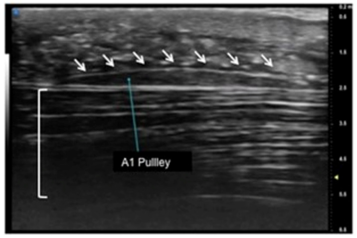
Sagittal view of A1 pulley (white arrows) presenting as a fusiform structure with a hypo-echoic signal contoured by a thin hyperechoic line. The superficial flexor tendon is visible (square parenthesis).
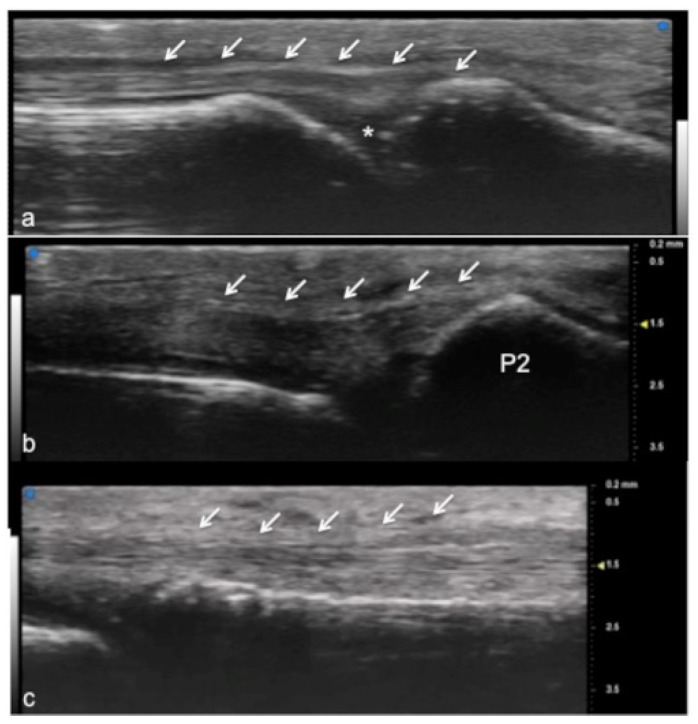
Extensor tendon of a finger. UHFUS gives a detailed and magnified representation of small structures such as the extensor tendon of the finger, even allowing the visualization of partial lesions. In (a), the sagittal view of the terminal extensor tendon (white arrows) at the level of the distal interphalangeal joint (white asterisk). In (b), the median band of the extensor tendon inserting at the level of the middle phalanx (P2). In (c), the thin sagittal band of the extensor tendon at the level of the metacarpal head.
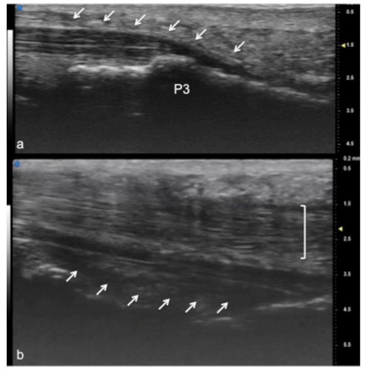
Flexor tendon of a finger. Using UHFUS at the flexor tendons, the same spatial resolution can be achieved as in the imaging of the extensor tendons. In (a), the sagittal view of the deep flexor tendon component inserting on the basis of the distal phalanx (P3). In (b), the superficial component of the flexor tendon (white arrows) lying near the deep component (white square parenthesis).
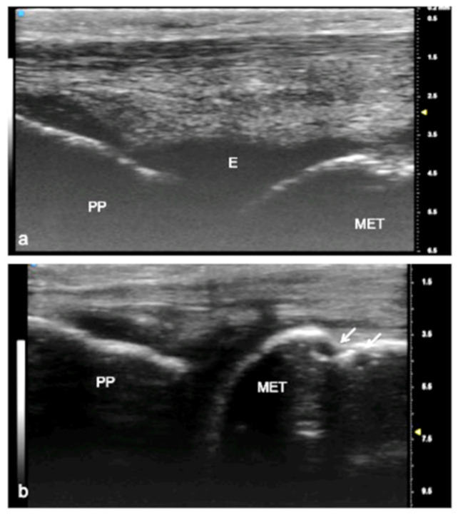
Juvenile idiopathic arthritis (JIA). UHFUS gives clear details of pathological findings in pediatric patients with JIA. In (a), articular effusion (E) at the level of the metacarpal–phalangeal joint (MET-PP). In (b), osseous erosions at the level of the metacarpal head (white arrows).
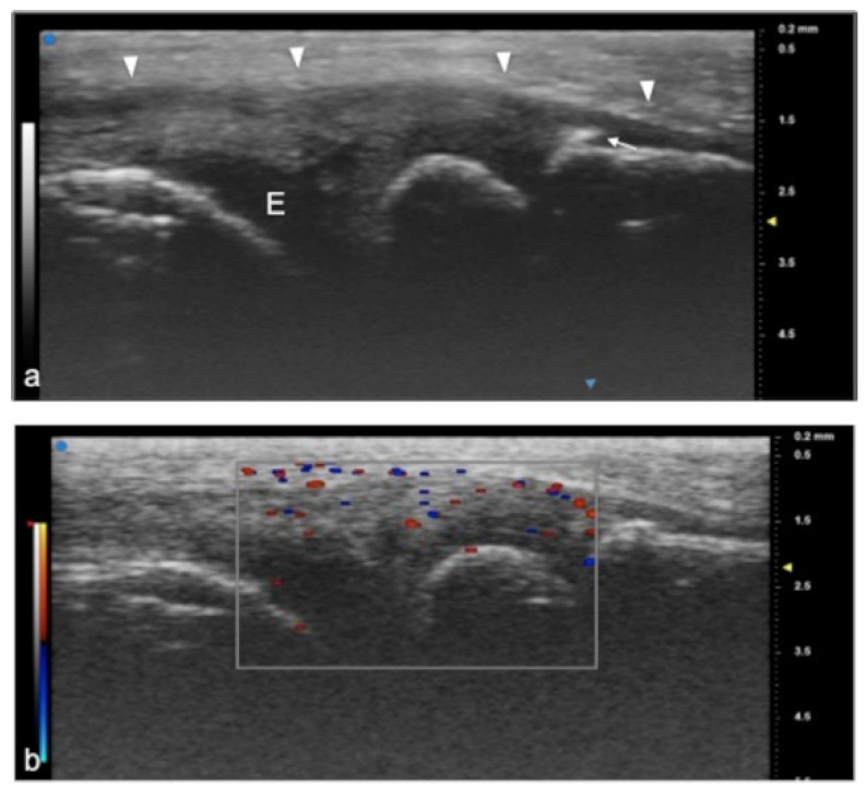
Juvenile idiopathic arthritis. UHFUS gives clear details of pathological findings in pediatric patients with JIA. In (a), articular effusion (E) at the level of wrist and enthesophyte (white arrow). Thickened capsule (arrowheads) in (a) presenting with an increased Doppler signal (white square) in (b).
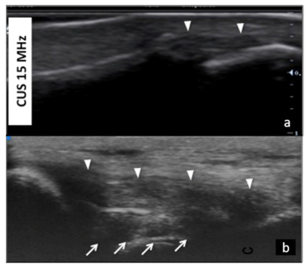
Collateral ligaments of interphalangeal joints: comparison between 50 MHz probes in (a) and 15 MHz CUS (arrowheads) in (b). In (a), UHFUS gives a more detailed and magnified representation of both deep (white arrows) and superficial (white arrowheads) components of the ligament.
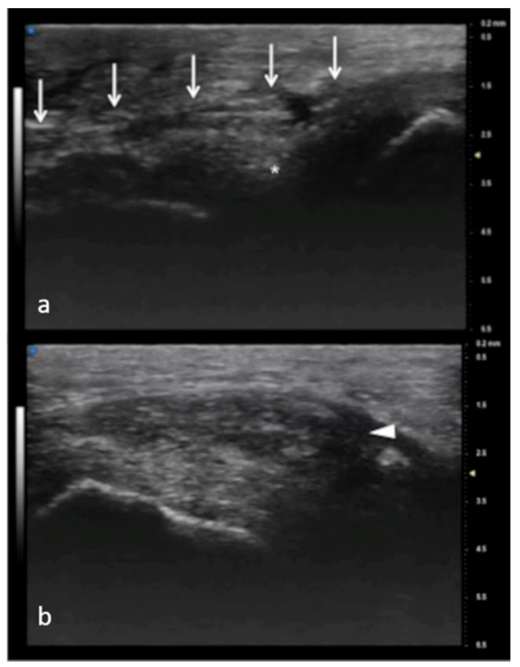
Full-thickness UCL tear. In (a), normal appearance of adductor pollicis aponeurosis (white arrows) and below the ulnar collateral ligament (asterisk) of the first metacarpophalangeal joint. In (b), the non-visualization of the ulnar collateral ligament and the presence of a mass-like area (arrowhead) proximal to the joint have high accuracy in depicting a displaced full-thickness ligament tear.
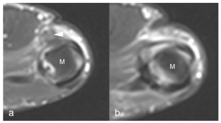
Same patient as in Figure 7. 1.5 T MRI shows a mass-like area (arrowhead) proximal to the joint representing the injured and displaced ligament in (a). In (b), the contralateral metacarpal phalangeal joint of the thumb. M: metacarpal head.
Visualization of small peripheral nerves using UHFUS. Recurrent branch (line) of the median nerve (square parenthesis) at the level of the thenar eminence in (a). Digital branch (dot circle) of the median nerve at the level of the finger in (b).
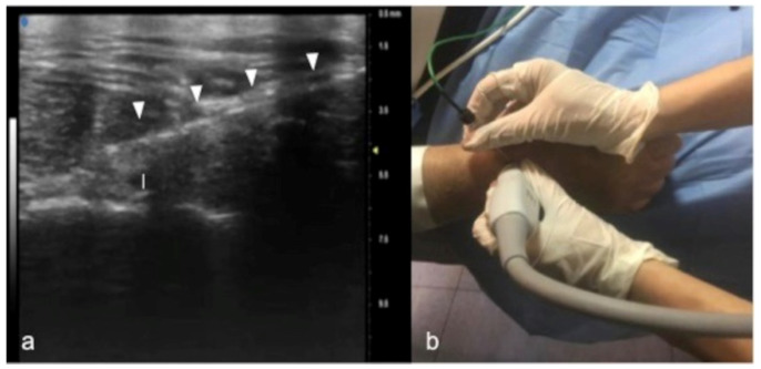
Percutaneous radiofrequency ablation of the posterior interosseous nerve for chronic wrist pain: In (a), cannula’s insertion (white arrowheads) under real-time ultrasound. Guidance for direct visualization of the posterior interosseous nerve (white caliber). The procedure is performed on an awake patient using a noninvasive approach (b).
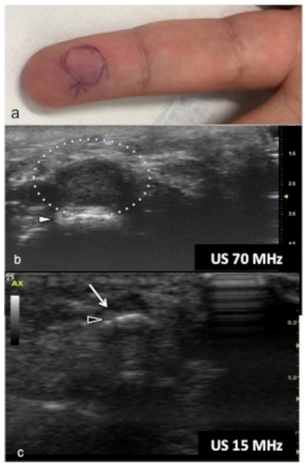
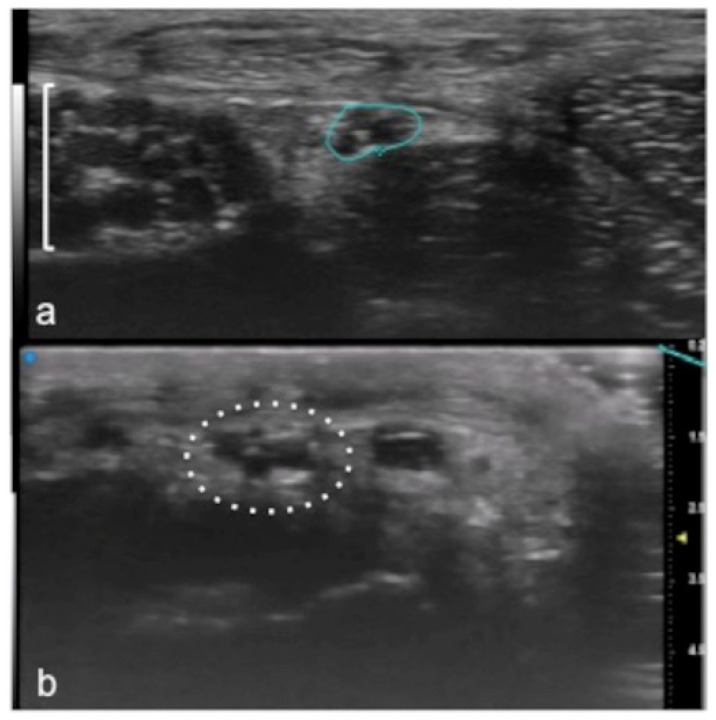
Glomus tumor. The location corresponds to the pulp of finger IV (a). The patient had the classical triad of symptoms: paroxysmal pain, pinpoint pain, and cold hypersensitivity, lasting for two years. In (b), UHFUS shows a well-delimited nodule in contact with the adjacent phalangeal bone (arrowhead), but no cortical deformity is present. No significant hyperemia on color Doppler was noticed. On CUS (c), the nodule was delineated only thanks to the help of the preliminary UHFUS exam.
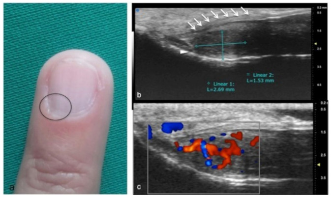
Glomus tumor in the subungual space. No clear alterations were visible during clinical examination at the site of pain (circle in (a)). In (b), UHFUS effectively demonstrates the presence of a hypo-isoechogenic nodule (calibers) in contact with the adjacent phalangeal bone (arrowhead) under the nail plate (arrows). Note the small deformation of the nail plate. Mild vascularization on color Doppler was present (c).
References
-
- Izzetti R., Vitali S., Aringhieri G., Nisi M., Oranges T., Dini V., Ferro F., Baldini C., Romanelli M., Caramella D., et al. Ultra-High Frequency Ultrasound, A Promising Diagnostic Technique: Review of the Literature and Single-Center Experience. Can. Assoc. Radiol. J. 2020;72:418–431. doi: 10.1177/0846537120940684. - DOI - PubMed
-
- Vogt M., Knüttel A., Hoffmann K., Altmeyer P., Ermert H. Comparison of High Frequency Ultrasound and Optical Coherence Tomography as Modalities for High Resolution and Non Invasive Skin Imaging. Vergleich von hochfrequentem Ultraschall und optischer Kohärenztomographie als Modalitäten für die hochauflösende und nichtinvasive Abbildung der Haut. Biomed. Eng./Biomed. Tech. 2003;48:116–121. doi: 10.1515/bmte.2003.48.5.116. - DOI - PubMed
Publication types
LinkOut - more resources
Full Text Sources
