. 2022 Sep 27;12(10):1500.
doi: 10.3390/life12101500.

Olive Leaf Extract Attenuates Chlorpyrifos-Induced Neuro- and Reproductive Toxicity in Male Albino Rats

Affiliations

Olive Leaf Extract Attenuates Chlorpyrifos-Induced Neuro- and Reproductive Toxicity in Male Albino Rats

Arwa A Hassan et al. Life (Basel). .

Abstract

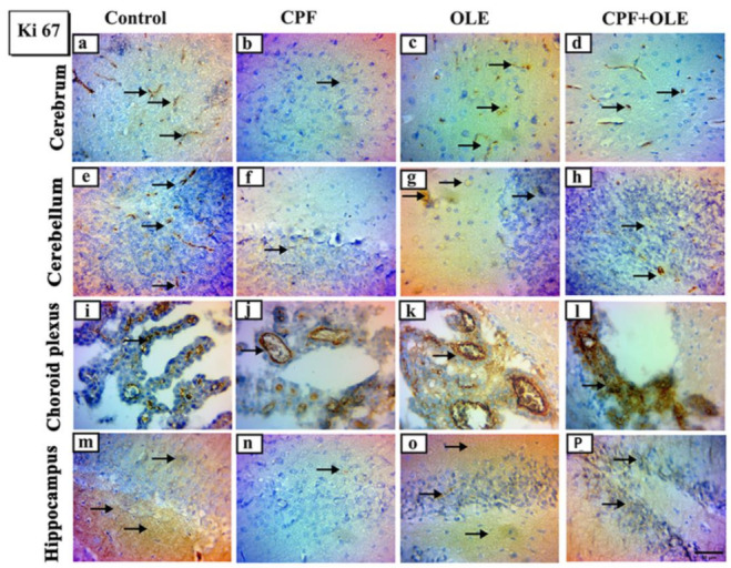
Chlorpyrifos (CPF) is a common organophosphorus insecticide. It is associated with negative consequences such as neurotoxicity and reproductive injury. This study aimed to observe the ability of olive leaf extract to attenuate chlorpyrifos toxicity, which induced neuro- and reproductive toxicity in male albino rats. Olive leaf extract (OLE) exhibits potent antioxidant and antiapoptotic properties. Twenty-two mature male rats were divided into four groups: control (saline), CPF (9 mg/kg), OLE (150 mg/kg), and CPF + OLE. Treatment was administered orally for 80 days. The CPF significantly reduced serum sex hormones, sperm counts and motility, high oxidants (MDA), and depleted antioxidants (GSH, SOD, TAC) in the brain and testes homogenate; additionally, it decreased serum AChE and brain neurotransmitters, increased Bax, decreased Bcl-2, and boosted caspase-3 immune expression in neural and testicular cells. Immunological expression of Ki 67 in the cerebrum, cerebellum, choroid plexus, and hippocampus was reduced, and α-SMA in testicular tissue also decreased. Histopathological findings were consistent with the above impacts. OLE co-administration significantly normalized all these abnormalities. OLE showed significant protection against neural and reproductive damage caused by CPF.

Keywords: apoptosis; chlorpyrifos; neurotoxicity; olive leaf extract; oxidative stress; reproductive toxicity.


Conflict of interest statement

The authors declare no conflict of interest.

Figures

Figure 1
Figure 1
Representative photomicrographs of hematoxylin-eosin and immunohistochemical stained cross-sections of the testicular rats, in control (a,f,o), CPF-treated (b,g,p), OLE-treated (c,h,q), and CPF + OLE-treated groups (d,i,r). The testicular architecture for (a,c,d) groups showed seminiferous tubules lined by spermatogonia (G), spermatocytes (C), spermatids (D), immature spermatozoa (IS), and Sertoli cells (S) surrounded by myoid cells (M) and Leydig cells (L) relative to the b group, which showed disorganized spermatogenic epithelium (closed arrows) and hyalinization (asterisks) with vacuolization (arrowheads) of the testicular interstitium (scale bar: 50 μm). Expressions of Myoid cells via α-SMA (fn) and apoptotic cell populations via caspase-3 protein (ow) immunostaining of testicular rats, Arrows indicate dark brown staining of positive immune cells. (jm) Moreover, (sv) show selected areas of the seminiferous epithelium at higher magnifications. Scale bar: (fi) and (or) 100 μm (jm); (sv) 50 μm. Bar charts demonstrating seminiferous epithelial height (e) and area % of α-SMA- and caspase-3-positive expressions (n and w, respectively) in the testicular sections of four experimental groups. Bars carrying different superscripts (a–c) are statistically significant differences at p ≤ 0.05, as determined using one-way ANOVA followed by Duncan’s test, n = 5/group. Values are expressed as the means ± SE.
Figure 2
Figure 2
Representative photomicrographs of hematoxylin and eosin-stained parasagittal sections of the brain of rats in control (a), CPF-treated (b), OLE-treated (c), and CPF + OLE-treated groups (d). A photomicrograph of the control and OLE-treated (a,b) groups shows a normally structured cerebrum having larger neuronal cell bodies with vesicular nuclei (arrows). CPF-treated (c) group showing dark, shrunken neuronal cell bodies with deeply stained pyknotic nuclei (arrowheads) and congested blood capillaries (crooked arrow). CPF + OLE-treated group (d) showing many normal cell bodies having vesicular nuclei (arrows), while a few neuronal cell bodies appear dark and shrunken with pyknotic nuclei (arrowheads). Photomicrograph of cerebellum of control and OLE-treated (a,b) groups showing normal structured cerebellar layers as outer molecular (OML), inner granular (IGL), and middle Purkinje cells (arrowheads) that are regularly located between the OML and IGL. CPF treated (c) group showing a few numbers of Purkinje cell bodies with the presence of some necrotic Purkinje cells (arrow) and congested blood capillaries (crooked arrow). The CPF + OLE-treated (d) group showing a disorganized alignment of the Purkinje cells (arrowheads). Photomicrograph of choroid plexus: control and OLE-treated (a,b) groups showing normally structured choroid plexus. CPF-treated (c) group showing blood capillary congestion (zigzag arrows) and proliferation of the lining epithelial (arrowhead). CPF + OLE-treated group (d) showing blood capillary congestion only (zigzag arrows). The photomicrograph of the hippocampus of the control (a) group has the dentate gyrus (DG) and cornu ammonis (CA1,2,3,4). Higher magnification of the squared area of the CA2 region (b): OLE-treated (C) groups showing well-defined molecular layer (ML), pyramidal layer (PL), and polymorphic layer (PML) with PL shows closely packed cell bodies of the pyramidal neurons (thick arrow). CPF-treated (c) group showing an apparent disruption in CA2 PL shows disorganized and loosely packed cell bodies (asterisk) that have pyknotic nuclei (arrowheads). The CPF + OLE-treated group (d) showing preserved layers with few pyknotic nuclei (arrowheads) and congested blood capillaries (crooked arrow). Scale bar: (ae) cerebrum, cerebellum, and choroid plexus, 50 μm (a); (be) the hippocampus, 200 μm and 100 μm.
Figure 3
Figure 3
Representative photomicrographs of Ki-67 immunohistochemical stained parasagittal sections of the brain of rats in control CPF, OLE and CPF+OLE treated groups, Arrows indicating dark brown staining of immune positive proliferating cells. Photomicrograph of cerebrum of: Control and OLE treated (a,c) groups showing a moderate positive reaction. CPF treated (b) group showing a weak positive reaction. CPF+OLE treated group (d) showing a mild positive reaction. Photomicrograph of cerebellum of: Control and OLE treated (e,g) groups showing a moderate positive reaction. CPF treated (f) group showing a weak positive reaction. CPF+OLE treated (h) group showing a mild positive reaction. Photomicrograph of choroid plexus of: Control and OLE treated (i,k) groups showing a moderate positive reaction. CPF treated (j) group showing a weak positive reaction. CPF+OLE treated group (l) showing a moderate positive reaction. Photomi-crograph of the hippocampus of: Control and OLE treated (m,o) groups showing a mild positive reaction. CPF treated (n) group showing a weak positive reaction. CPF+OLE treated group (p) showing a moderate positive reaction. Scale bar; 50 μm.
Figure 4
Figure 4
Representative photomicrographs of caspase-3 immunohistochemical stained parasagittal sections of the brain of rats in control, CPF, OLE and CPF+OLE treated groups, Arrows in-dicating dark brown staining of immune positive apoptotic cells. Photomicrograph of cerebrum of: Control and OLE treated (a,c) groups showing a weak positive reaction. CPF treated (b) group showing an intense positive reaction. CPF+OLE treated group (d) showing a mild positive reaction. Photomicrograph of cerebellum of: Control and OLE treated (e,g) groups showing a weak posi-tive reaction. CPF treated (f) group showing an intense positive reaction. CPF+OLE treated (h) group showing a mild positive reaction. Photomicrograph of choroid plexus of: Control and OLE treated (i,k) groups showing a weak positive reaction. CPF treated (j) group showing an intense positive reaction. CPF+OLE treated group (l) showing a mild positive reaction. Photomicrograph of the hippocampus of: Control and OLE treated (m,o) groups showing a weak positive reaction. CPF treated (n) group showing an intense positive reaction. CPF+OLE treated group (p) showing a mild positive reaction. Scale bar; 50 μm.

References

    1. Khalaf A., Ibrahim M., Tohamy A., Allah A., Zaki A.R. Protective effect of vitazinc on chlorsan induced oxidative stress, genotoxicity and histopathological changes in testicular tissues of male rats. Int. J. Pharmacol. 2017;13:22–32. doi: 10.3923/ijp.2017.22.32. - DOI
    1. Morgan A.M., Ibrahim M.A., Noshy P.A. Reproductive toxicity provoked by titanium dioxide nanoparticles and the ameliorative role of Tiron in adult male rats. Biochem. Biophys. Res. Commun. 2017;486:595–600. doi: 10.1016/j.bbrc.2017.03.098. - DOI - PubMed
    1. Shou L., Bei Y., Song Y., Wang L., Ai L., Yan Q., He W. Nrf2 mediates the protective effect of edaravone after chlorpyrifos-induced nervous system toxicity. Environ. Toxicol. 2019;34:626–633. doi: 10.1002/tox.22728. - DOI - PubMed
    1. Mahmoud S.M., Abdel Moneim A.E., Qayed M.M., El-Yamany N.A. Potential role of N-acetylcysteine on chlorpyrifos-induced neurotoxicity in rats. Environ. Sci. Pollut. Res. 2019;26:20731–20741. doi: 10.1007/s11356-019-05366-w. - DOI - PubMed
    1. Singh N., Lawana V., Luo J., Phong P., Abdalla A., Palanisamy B., Rokad D., Sarkar S., Jin H., Anantharam V. Organophosphate pesticide chlorpyrifos impairs STAT1 signaling to induce dopaminergic neurotoxicity: Implications for mitochondria mediated oxidative stress signaling events. Neurobiol. Dis. 2018;117:82–113. doi: 10.1016/j.nbd.2018.05.019. - DOI - PMC - PubMed