. 2022 Oct 1;12(10):1534.
doi: 10.3390/life12101534.

Confocal Raman Micro-Spectroscopy for Discrimination of Glycerol Diffusivity in Ex Vivo Porcine Dura Mater

Affiliations

Confocal Raman Micro-Spectroscopy for Discrimination of Glycerol Diffusivity in Ex Vivo Porcine Dura Mater

Ali Jaafar et al. Life (Basel). .

Abstract

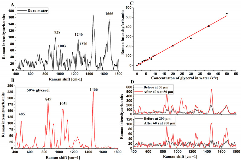
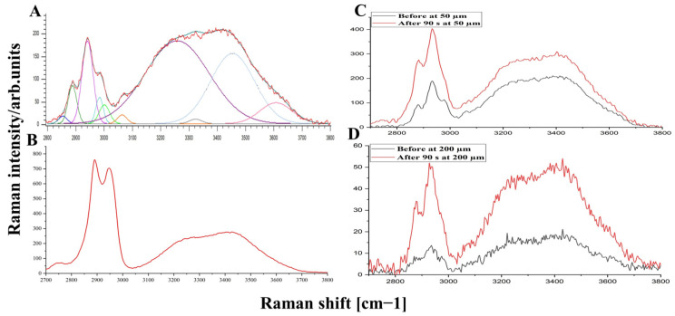
Dura mater (DM) is a connective tissue with dense collagen, which is a protective membrane surrounding the human brain. The optical clearing (OC) method was used to make DM more transparent, thereby allowing to increase in-depth investigation by confocal Raman micro-spectroscopy and estimate the diffusivity of 50% glycerol and water migration. Glycerol concentration was obtained, and the diffusion coefficient was calculated, which ranged from 9.6 × 10⁻⁶ to 3.0 × 10⁻⁵ cm2/s. Collagen-related Raman band intensities were significantly increased for all depths from 50 to 200 µm after treatment. In addition, the changes in water content during OC showed that 50% glycerol induces tissue dehydration. Weakly and strongly bound water types were found to be most concentrated, playing a major role in the glycerol-induced water flux and OC. Results show that OC is an efficient method for controlling the DM optical properties, thereby enhancing the in-depth probing for laser therapy and diagnostics of the brain. DM is a comparable to various collagen-containing tissues and organs, such as sclera of eyes and skin dermis.

Keywords: collagen type I; dehydration; diffusion coefficients; glycerol; high wavenumber; hydrogen bound water; penetration; topical application.


Conflict of interest statement

The authors declare that there are no conflict of interest.

Figures

Figure 1
Figure 1
Schematic illustration of the measurement of DM samples with CRM.
Figure 2
Figure 2
FP Raman spectra of control porcine DM at 50 µm depth (A), 50% glycerol (B), calibration curve of Raman band intensity at 485 cm−1, and concentration of glycerol solution in 0–50% water (C). The red line (in C) is a result of fitting with correlation coefficient (R2) of 0.9941 and the average Raman spectra before and after 60 s treatment at 50 and 200 µm depths (D).
Figure 3
Figure 3
The time dependencies of DM collagen-related Raman bands at 938 (black squares), 1003 (red circles), 1247 (blue upward triangles), 1270 (green downward triangles), and 1665 cm−1 (purple diamonds) after impregnation by 50% aqueous-glycerol solution from 0 to 390 s at depths 50 (A), 100 (B), 150 (C), and 200 µm (D).
Figure 4
Figure 4
Gaussian lines function-based deconvolution of the HWN Raman spectrum of untreated porcine DM at 50 μm (A), 50% glycerol water solution (B), and the average of Raman spectra before and after 90 s treatment at 50 and 200 μm depths (C and D), respectively.
Figure 5
Figure 5
The time-dependent kinetics of 3005 cm−1 (tightly bound water, black), 3270 cm−1 (strongly bound water, red), 3458 cm−1 (weakly bound water, blue), and 3605 cm−1 (unbound water, green) deconvoluted Gaussian lines at depths of 50 (A), 100 (B), 150 (C), and 200 µm (D).
Figure 6
Figure 6
The kinetics of total water content changes in 50% glycerol-treated porcine DM at depths: 50 (black), 100 (red), 150 (blue), and 200 µm (green) depending on treatment time.

References

    1. Latka I., Dochow S., Krafft C., Dietzek B., Popp J. Fiber optic probes for linear and nonlinear Raman applications-Current trends and future development. Laser Photonics Rev. 2013;7:698–731. doi: 10.1002/lpor.201200049. - DOI
    1. Darvin M.E., Schleusener J., Lademann J., Choe C.-S. Current views on non-invasive in vivo determination of physiological parameters of the stratum corneum using confocal Raman microspectroscopy. Skin Pharmacol. Physiol. 2022;35:125–136. doi: 10.1159/000521416. - DOI - PubMed
    1. DePaoli D., Lemoine É., Ember K., Parent M., Prud’homme M., Cantin L., Petrecca K., Leblond F., Côté D.C. Rise of Raman spectroscopy in neurosurgery: A review. J. Biomed. Opt. 2020;25:1. doi: 10.1117/1.JBO.25.5.050901. - DOI - PMC - PubMed
    1. Stevens A.R., Stickland C.A., Harris G., Ahmed Z., Oppenheimer P.G., Belli A., Davies D.J. Raman Spectroscopy as a Neuromonitoring Tool in Traumatic Brain Injury: A Systematic Review and Clinical Perspectives. Cells. 2022;11:1227. doi: 10.3390/cells11071227. - DOI - PMC - PubMed
    1. Lee K.S., Landry Z., Pereira F.C., Wagner M., Berry D., Huang W.E., Taylor G.T., Kneipp J., Popp J., Zhang M., et al. Raman microspectroscopy for microbiology. Nat. Rev. Methods Prim. 2021;1:1–25. doi: 10.1038/s43586-021-00075-6. - DOI

LinkOut - more resources