Exosomes Derived from Bone Marrow Mesenchymal Stem Cells Alleviate Ischemia-Reperfusion Injury and Promote Survival of Skin Flaps in Rats
- PMID: 36295004
- PMCID: PMC9604753
- DOI: 10.3390/life12101567
Exosomes Derived from Bone Marrow Mesenchymal Stem Cells Alleviate Ischemia-Reperfusion Injury and Promote Survival of Skin Flaps in Rats
Abstract
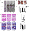
Free tissue flap transplantation is a classic and important method for the clinical repair of tissue defects. However, ischemia-reperfusion (IR) injury can affect the success rate of skin flap transplantation. We used a free abdomen flap rat model to explore the protective effects of exosomes derived from bone marrow mesenchymal stem cells (BMSCs-exosomes) against the IR injury of the skin flap. Exosomes were injected through the tail vein and the flaps were observed and obtained on day 7. We observed that BMSCs-exosomes significantly reduced the necrotic lesions of the skin flap. Furthermore, BMSCs-exosomes relieved oxidative stress and reduced the levels of inflammatory factors. Apoptosis was evaluated via the terminal deoxynucleotidyl transferase dUTP nick-end labeling (TUNEL) assay and Western blot analysis of Bax, Bcl-2. Simultaneously, BMSCs-exosomes promoted the formation of new blood vessels in the IR flap, as confirmed by the increased expression level of VEGFA and the fluorescence co-staining of CD31 and PCNA. Additionally, BMSCs-exosomes considerably increased proliferation and migration of human umbilical vein endothelial cells and promoted angiogenesis in vitro. BMSCs-exosomes could be a promising cell-free therapeutic candidate to reduce IR injury and promote the survival of skin flaps.
Keywords: angiogenesis; bone marrow mesenchymal stem cells; exosomes; ischemia-reperfusion injury; skin flap.
Conflict of interest statement
The authors declare no conflict of interest.
Figures


Similar articles
-
Hypoxic mesenchymal stem cell-derived exosomes promote the survival of skin flaps after ischaemia-reperfusion injury via mTOR/ULK1/FUNDC1 pathways.J Nanobiotechnology. 2023 Sep 21;21(1):340. doi: 10.1186/s12951-023-02098-5. J Nanobiotechnology. 2023. PMID: 37735391 Free PMC article.
-
Exosomes in Skin Flap Survival: Unlocking Their Role in Angiogenesis and Tissue Regeneration.Biomedicines. 2025 Feb 4;13(2):353. doi: 10.3390/biomedicines13020353. Biomedicines. 2025. PMID: 40002766 Free PMC article. Review.
-
Adipose mesenchymal stem cell-derived exosomes stimulated by hydrogen peroxide enhanced skin flap recovery in ischemia-reperfusion injury.Biochem Biophys Res Commun. 2018 Jun 2;500(2):310-317. doi: 10.1016/j.bbrc.2018.04.065. Epub 2018 Apr 14. Biochem Biophys Res Commun. 2018. PMID: 29654765
-
Bone Marrow Mesenchymal Stem Cells-derived Exosomes Promote Survival of Random Flaps in Rats through Nrf2-mediated Antioxidative Stress.J Reconstr Microsurg. 2025 Mar;41(3):177-190. doi: 10.1055/a-2331-8046. Epub 2024 May 23. J Reconstr Microsurg. 2025. PMID: 38782030
-
Multifunctional exosomes derived from bone marrow stem cells for fulfilled osseointegration.Front Chem. 2022 Aug 22;10:984131. doi: 10.3389/fchem.2022.984131. eCollection 2022. Front Chem. 2022. PMID: 36072705 Free PMC article. Review.
Cited by
-
Hypoxia in Skin Cancer: Molecular Basis and Clinical Implications.Int J Mol Sci. 2023 Feb 23;24(5):4430. doi: 10.3390/ijms24054430. Int J Mol Sci. 2023. PMID: 36901857 Free PMC article. Review.
-
Hypoxic mesenchymal stem cell-derived exosomes promote the survival of skin flaps after ischaemia-reperfusion injury via mTOR/ULK1/FUNDC1 pathways.J Nanobiotechnology. 2023 Sep 21;21(1):340. doi: 10.1186/s12951-023-02098-5. J Nanobiotechnology. 2023. PMID: 37735391 Free PMC article.
-
Engineering exosomes from fibroblast growth factor 1 pre-conditioned adipose-derived stem cells promote ischemic skin flaps survival by activating autophagy.Mater Today Bio. 2024 Oct 26;29:101314. doi: 10.1016/j.mtbio.2024.101314. eCollection 2024 Dec. Mater Today Bio. 2024. PMID: 39534677 Free PMC article.
-
Exosomes in Skin Flap Survival: Unlocking Their Role in Angiogenesis and Tissue Regeneration.Biomedicines. 2025 Feb 4;13(2):353. doi: 10.3390/biomedicines13020353. Biomedicines. 2025. PMID: 40002766 Free PMC article. Review.
-
The Protective Effects of MSC-Derived Exosomes Against Chemotherapy-Induced Parotid Gland Cytotoxicity.Int J Dent. 2025 Apr 3;2025:5517092. doi: 10.1155/ijod/5517092. eCollection 2025. Int J Dent. 2025. PMID: 40223864 Free PMC article.
References
-
- Dort J.C., Farwell D.G., Findlay M., Huber G.F., Kerr P., Shea-Budgell M., Simon C., Uppington J., Zygun D., Ljungqvist O., et al. Optimal Perioperative Care in Major Head and Neck Cancer Surgery with Free Flap Reconstruction: A Consensus Review and Recommendations from the Enhanced Recovery After Surgery Society. JAMA Otolaryngol. Neck Surg. 2017;143:292–303. doi: 10.1001/jamaoto.2016.2981. - DOI - PubMed
-
- Akdeniz-Dogan Z., Roubaud M.S., Kapur S.K., Liu J., Yu P., Selber J.C., Mericli A.F. Free Flap Reconstruction of Posterior Trunk Soft Tissue Defects: Single-Institution Experience and Systematic Literature Review. Plast. Reconstr. Surg. 2021;147:728–740. doi: 10.1097/PRS.0000000000007675. - DOI - PubMed
-
- Odake K., Tsujii M., Iino T., Chiba K., Kataoka T., Sudo A. Febuxostat treatment attenuates oxidative stress and inflammation due to ischemia-reperfusion injury through the necrotic pathway in skin flap of animal model. Free Radic. Biol. Med. 2021;177:238–246. doi: 10.1016/j.freeradbiomed.2021.10.033. - DOI - PubMed
Grants and funding
- 81974144/National Natural Science Foundation of China
- 82072984/National Natural Science Foundation of China
- Z161100000516201/the Beijing Municipal Science and Technology Commission
- YSP202008/Beijing Stomatological Hospital, Capital Medical University Young Scientist Program
- 18-09-21/the Discipline Construction Fund of Beijing Stomatological Hospital
LinkOut - more resources
Full Text Sources
Research Materials
Miscellaneous

