Review
. 2022 Oct 14;12(10):1601.
doi: 10.3390/life12101601.

Single Cell in a Gravity Field

Affiliations
Review

Single Cell in a Gravity Field

Irina V Ogneva. Life (Basel). .

Abstract

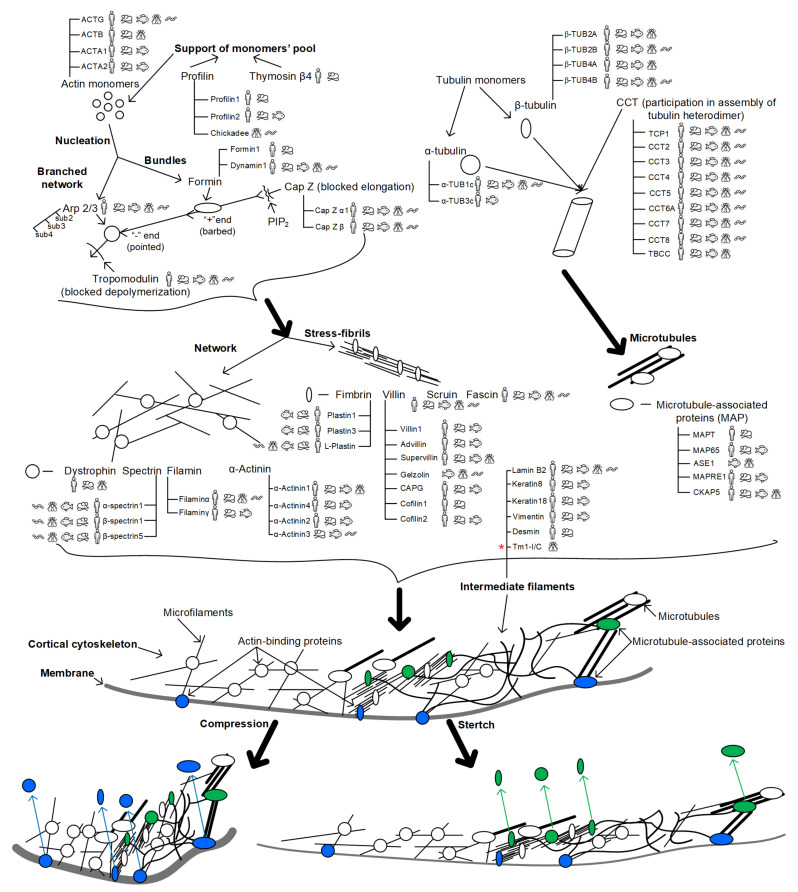
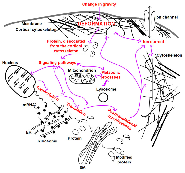
The exploration of deep space or other bodies of the solar system, associated with a long stay in microgravity or altered gravity, requires the development of fundamentally new methods of protecting the human body. Most of the negative changes in micro- or hypergravity occur at the cellular level; however, the mechanism of reception of the altered gravity and transduction of this signal, leading to the formation of an adaptive pattern of the cell, is still poorly understood. At the same time, most of the negative changes that occur in early embryos when the force of gravity changes almost disappear by the time the new organism is born. This review is devoted to the responses of early embryos and stem cells, as well as terminally differentiated germ cells, to changes in gravity. An attempt was made to generalize the data presented in the literature and propose a possible unified mechanism for the reception by a single cell of an increase and decrease in gravity based on various deformations of the cortical cytoskeleton.

Keywords: cellular mechanoreception; cellular mechanotransduction; early embryo; hypergravity; microgravity; oocyte; space flight; spermatozoon.


Conflict of interest statement

The author declares no conflict of interest.

Figures

Figure 1
Figure 1
Possible scheme of cellular mechanoreception. To demonstrate the variability of possible participants in mechanoreception, the main proteins involved in the organization of the components of the cytoskeleton are presented. Pictograms indicate the expression of these genes in various animal species—H. sapiens, M. musculus, D. rerio, D. melanogaster, and C. elegans (according to the open resource HomoloGene https://www.ncbi.nlm.nih.gov/homologene, accessed on 21 September 2022). Microfilaments: the pool of actin monomers is maintained by profilin family proteins and thymosin β4; monomers polymerize into filaments, and their length is controlled by tropomodulin (at the pointed end) and CapZ (at the barbed end); microfilaments either stack in bundles (with formin nucleation) or form a branched network (with Arp 2/3 nucleation); a network of microfilaments and stress fibrils is organized by actin-binding proteins. Microtubules: tubulin monomers, alpha- and beta-, form a heterodimer with the participation of proteins of the CCT family; heterodimers are assembled into microtubules, the spatial organization of which and association with other intracellular structures is carried out by MAP proteins. Intermediate filaments: due to the presence of rod-like domains in the monomers, intermediate filaments are assembled, which can be localized in the nucleus (lamins) and in the cytoplasm. Not so long ago, it was believed that Drosophila lacks cytoplasmic intermediate filaments [124] and that the cell structure is strengthened at the expense of other components of the cytoskeleton. Therefore, it seems important to note (by red asterisk) recent data indicating that D. melanogaster has cytoplasmic intermediate filaments formed by the Tm1-I/C protein [125,126]. A change in external mechanical stress (for example, gravity) will lead to deformation. Compressive deformation would possibly lead to dissociation from the cortical cytoskeleton of the proteins anchoring it to the membrane—these are highlighted in green. Tensile deformation may lead to dissociation from the cortical cytoskeleton of proteins that organize the parallel stacked components of the cytoskeleton—they are highlighted in blue. Highlighted proteins diffuse from the cortical cytoskeleton under tension and contraction, as indicated by colored arrows. In both cases, the choice of specific participants in the process can be species-specific.
Figure 2
Figure 2
Relationship of mechanotransduction pathways in a single cell. The main cellular structures are schematically indicated: membrane, cytoskeleton, nucleus, endoplasmic reticulum (ER) and ribosomes, Golgi apparatus (GA), mitochondrion, and lysosome. Cytoskeletal structures penetrate the cell through and through and connect all organelles to each other, forming a cytoskeletal network. The red labels indicate the main processes that can be targeted as a result of gravity change transduction. Purple arrows indicate possible mutual regulation of intracellular processes.

Similar articles

Cited by

References

    1. Ohira T., Kawano F., Goto K., Kaji H., Ohira Y. Responses of neuromuscular properties to unloading and potential countermeasures during space exploration missions. Neurosci. Biobehav. Rev. 2022;136:104617. doi: 10.1016/j.neubiorev.2022.104617. - DOI - PubMed
    1. Lee P.H.U., Chung M., Ren Z., Mair D.B., Kim D.-H. Factors mediating spaceflight-induced skeletal muscle atrophy. Am. J. Physiol.-Cell Physiol. 2022;322:C567–C580. doi: 10.1152/ajpcell.00203.2021. - DOI - PubMed
    1. Ohira T., Kawano F., Ohira T., Goto K., Ohira Y. Responses of skeletal muscles to gravitational unloading and/or reloading. J. Physiol. Sci. 2015;65:293–310. doi: 10.1007/s12576-015-0375-6. - DOI - PMC - PubMed
    1. Oganov V.S. Modern analysis of bone loss mechanisms in microgravity. J. Gravit. Physiol. 2004;11:143–146. - PubMed
    1. Vico L., Hargens A. Skeletal changes during and after spaceflight. Nat. Rev. Rheumatol. 2018;14:229–245. doi: 10.1038/nrrheum.2018.37. - DOI - PubMed

LinkOut - more resources