. 2022 Oct 17;15(20):7259.
doi: 10.3390/ma15207259.

Highlighting Bacteria with Calcifying Abilities Suitable to Improve Mortar Properties

Affiliations

Highlighting Bacteria with Calcifying Abilities Suitable to Improve Mortar Properties

Iuliana Răut et al. Materials (Basel). .

Abstract

Biomineralization, the use of microorganisms to produce calcium carbonate, became a green solution for application in construction materials to improve their strength and durability. The calcifying abilities of several bacteria were investigated by culturing on a medium with urea and calcium ions. The characterization of the precipitates from bacterial cultures was performed using X-ray diffraction, Fourier transform infrared spectroscopy, and thermogravimetric analysis. The formation of carbonate crystals was demonstrated by optical and scanning electron microscopy. Water absorption and compressive strength measurements were applied to mortars embedded with sporal suspension. The efficiency of the supplementation of mortar mixtures with bacterial cells was evaluated by properties, namely the compressive strength and the water absorption, which are in a relationship of direct dependence, the increase in compressive strength implying the decrease in water absorption. The results showed that Bacillus subtilis was the best-performing bacterium, its introduction into the mortar producing an increase in compressive strength by 11.81% and 9.50%, and a decrease in water absorption by 11.79% and 10.94%, after 28 and 56 days of curing, respectively, as compared to standards. The exploitation of B. subtilis as a calcifying agent can be an interesting prospect in construction materials.

Keywords: Bacillus; biomineralization; concrete; ureolytic bacteria.


Conflict of interest statement

The authors declare no conflict of interest.

Figures
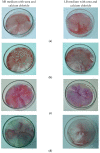
Figure 1
Figure 1
Alizarin colorimetric assay for testing carbonatogenesis ability of bacterial strains: (a) Bacillus amyloliquefaciens; (b) Bacillus licheniformis; (c) Bacillus subtilis; (d) Psuedomonas putida.
Figure 2
Figure 2
Optical microscopy applied to bacterial culture broth after coloring with red alizarin S (×40): (a) Bacillus amyloliquefaciens; (b) Bacillus subtilis; (c) Bacillus licheniformis. Black arrow—calcium carbonate crystals.
Figure 3
Figure 3
Ureolytic activity of bacterial strains in Petri plates on solid media of different compositions: (a) B. licheniformis on media M1, M2 and their corresponding controls (without urea and calcium ions); (b) B. subtilis on media M1, M2 and their corresponding controls (without urea and calcium ions); (c) B. amyloliquefaciens on media M1, M2 and their corresponding controls (without urea and calcium ions).
Figure 4
Figure 4
Images of experiments performed with Bacillus strains in medium with or without urea and Ca2+: (a) B. licheniformis; (b) B. subtilis; (c) B. amyloliquefaciens. In order from left to right, bacterial culture on TSB medium; bacterial culture on medium with urea and Ca2+; Mmedium, control, as medium mineral with urea and Ca2+, non-inoculated; Mmedium, control as medium mineral without urea and Ca2+, non-inoculated; red arrow—white deposit containing bacterial biomass and calcium carbonate.
Figure 5
Figure 5
Microscopic investigations of deposits obtained from bacteria cultured on medium with urea and calcium ions. On left, optical microscopy is shown, and on the right, SEM analysis, as: (a) Spores and crystals carbonate from Bacillus amyloliquefaciens (20×); (b) SEM analysis of pellet from Bacillus amyloliquefaciens (250×); (c) Spores and crystals carbonate from Bacillus licheniformis (20×); (d) SEM analysis of pellet from Bacillus licheniformis (250×); (e) Crystals carbonate and germens of crystallization at Bacillus subtilis (100×); (f) SEM analysis of deposit from Bacillus subtilis (5000×). Dotted square-bacterial cells; arrow—calcium carbonate crystals.
Figure 6
Figure 6
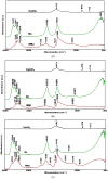
Overlaps of FTIR spectra of bacterial deposits from cultures on media with or without urea and calcium ions: (a) Bacillus amyloliquefaciens (b) Bacillus licheniformis; (c) Bacillus subtilis.
Figure 7
Figure 7
TGA curves of precipitates obtained from bacterial cultures on medium with or without urea and Ca2+: (a) TGA curve of precipitates obtained from B. amyloliquefaciens cultured on medium with (BA, left) or without urea and Ca2+ (MBA, right); (b) TGA curve of precipitates obtained from B. licheniformis cultured on medium with (BL, left) without urea and Ca2+ (MBL, right); (c) TGA curve of precipitates obtained from B. subtilis cultured on medium with (BS, left) or without urea and Ca2+ (MBS, right).
Figure 7
Figure 7
TGA curves of precipitates obtained from bacterial cultures on medium with or without urea and Ca2+: (a) TGA curve of precipitates obtained from B. amyloliquefaciens cultured on medium with (BA, left) or without urea and Ca2+ (MBA, right); (b) TGA curve of precipitates obtained from B. licheniformis cultured on medium with (BL, left) without urea and Ca2+ (MBL, right); (c) TGA curve of precipitates obtained from B. subtilis cultured on medium with (BS, left) or without urea and Ca2+ (MBS, right).
Figure 8
Figure 8
XRD patterns of deposits recovered from bacterial cultured on medium with or without urea and Ca2+ ions: (a) Bacillus licheniformis; (b) Bacillus subtilis; (c) Bacillus amyloliquefaciens (red line—sample; black line—control, medium without urea and Ca2+ ions; C—calcium carbonate as calcite).
Figure 8
Figure 8
XRD patterns of deposits recovered from bacterial cultured on medium with or without urea and Ca2+ ions: (a) Bacillus licheniformis; (b) Bacillus subtilis; (c) Bacillus amyloliquefaciens (red line—sample; black line—control, medium without urea and Ca2+ ions; C—calcium carbonate as calcite).
Figure 9
Figure 9
Compressive strength (a) and Water absorption (b) of mortar specimens after 2, 7, 28 and 56 days of curing.

Similar articles

References

    1. Han P., Geng W.J., Li M.N., Jia S.R., Yin J.L., Xu X.R. Improvement of Biomineralization of Sporosarcina pasteurii as Biocementing Material for Concrete Repair by Atmospheric and Room Temperature Plasma Mutagenesis and Response Surface Methodology. J. Microbiol. Biotechnol. 2021;31:1311–1322. doi: 10.4014/jmb.2104.04019. - DOI - PMC - PubMed
    1. Li M., Zhu X., Mukherjee A., Huang M., Achal V. Biomineralization in metakaolin modified cement mortar to improve its strength with lowered cement content. J. Hazard. Mater. 2017;329:178–184. doi: 10.1016/j.jhazmat.2017.01.035. - DOI - PubMed
    1. Seifan M., Berenjian A. Application of microbially induced calcium carbonate precipitation in designing bio self-healing concrete. World J. Microbiol. Biotechnol. 2018;34:168. doi: 10.1007/s11274-018-2552-2. - DOI - PubMed
    1. Shaheen N., Jalil A., Adnan F., Khushnood R.A. Isolation of alkaliphilic calcifying bacteria and their feasibility for enhanced CaCO3 precipitation in bio-based cementitious composites. Microb. Biotechnol. 2021;14:1044–1059. doi: 10.1111/1751-7915.13752. - DOI - PMC - PubMed
    1. Lee Y.S., Park W. Current challenges and future directions for bacterial self-healing concrete. Appl. Microbiol. Biotechnol. 2018;102:3059–3070. doi: 10.1007/s00253-018-8830-y. - DOI - PubMed

LinkOut - more resources