

Transcallosal and Pericallosal Courses of the Anterior Cerebral Artery
- PMID: 36295526
- PMCID: PMC9608487
- DOI: 10.3390/medicina58101365
Transcallosal and Pericallosal Courses of the Anterior Cerebral Artery
Abstract
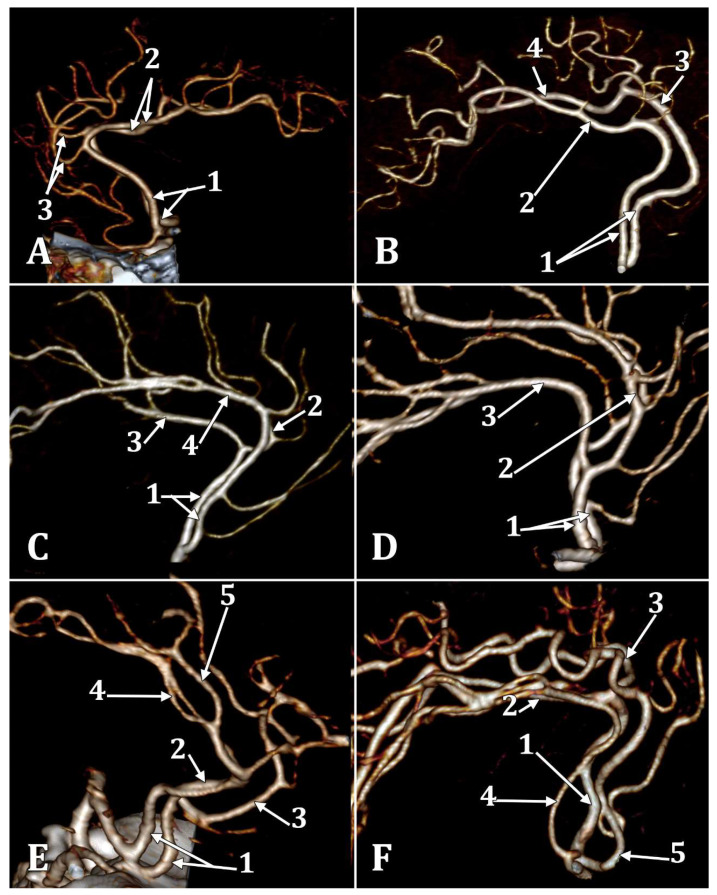
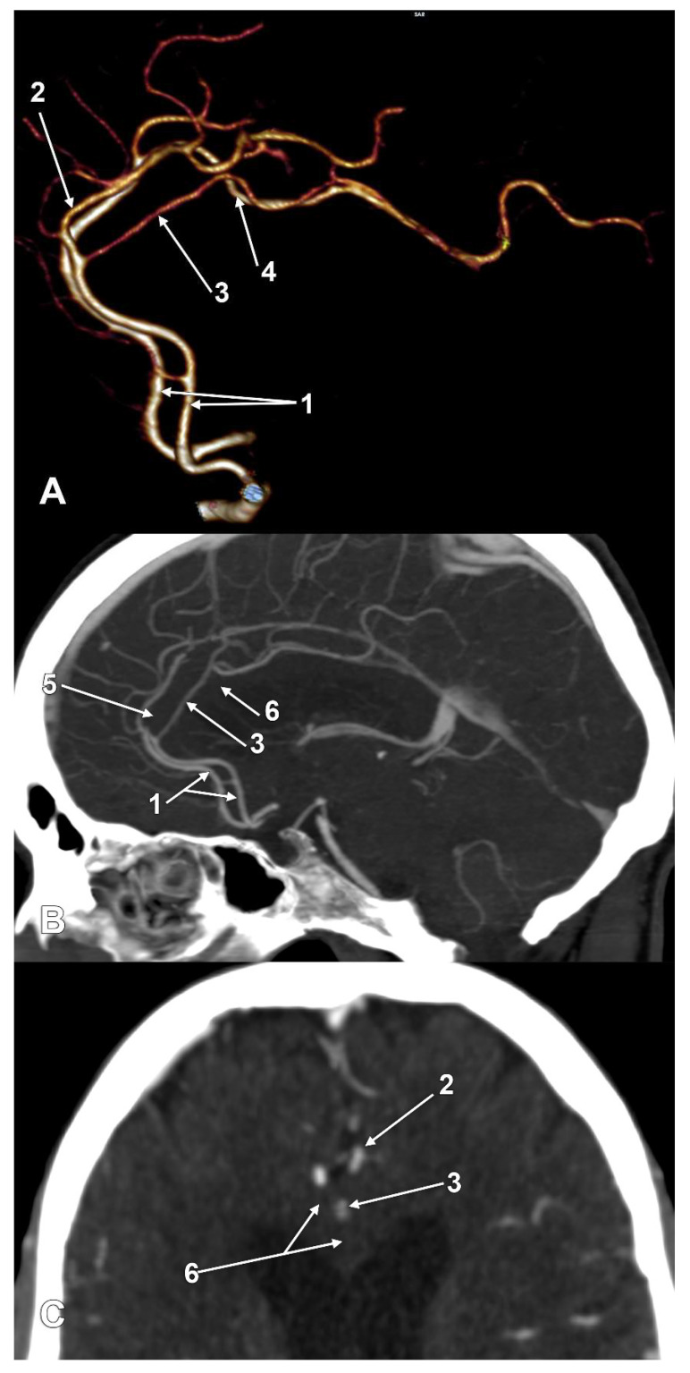
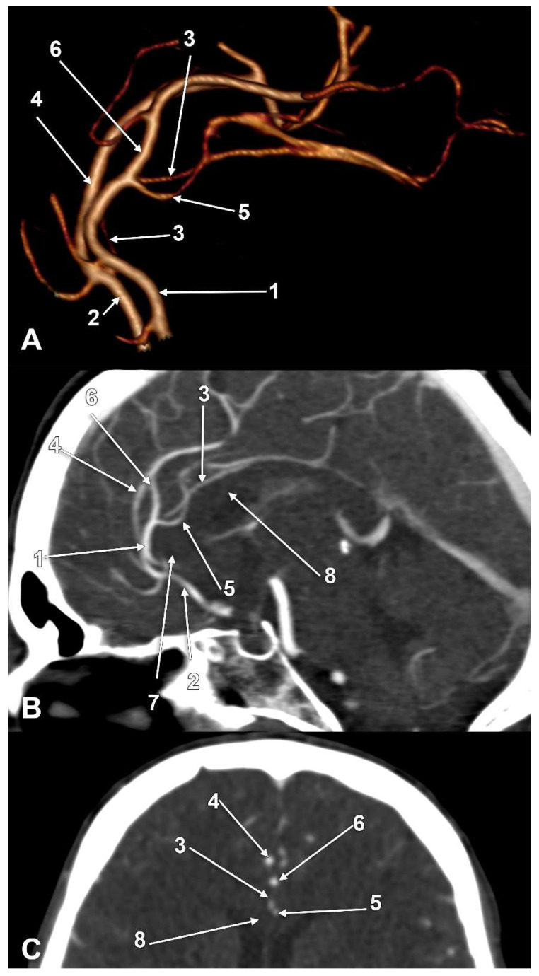
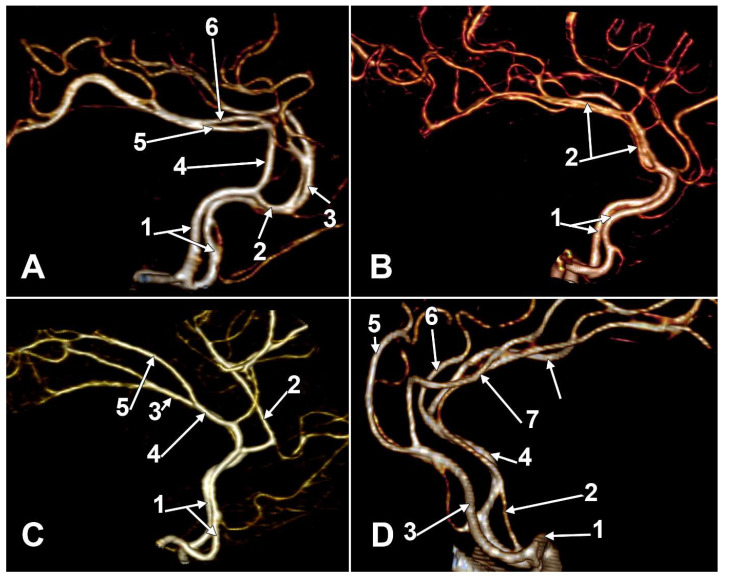
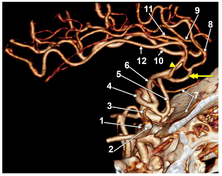
(1) Background: The anterior cerebral artery (ACA) has a precommunicating A1 segment, followed by a postcommunicating A2 segment. Anatomically, after it sends off from the callosomarginal artery (CMA), it continues as the pericallosal artery (PCalA). A detailed pattern of the anatomical variations of the PCalA are needed for practical reasons. (2) Methods: There were 45 retrospectively documented Computed Tomography Angiograms of 32 males and 13 females. (3) Results: In 90 sides, eleven different types of PCalA were documented: type 1: normal origin, above the genu of the corpus callosum (CC) (51.11%); type 2: low origin, below the rostrum of the CC (8.88%); type 3: late origin, above the body of the CC (3.33%); type 4, initial transcallosal course (3.33%); type 5, duplicated PCalA (1.11%); type 6, azygos PCalA (2.22%); type 7, absent PCalA (CMA type of ACA) (7.78%); type 8: CMA continued as PCalA (5.56%); type 9: PCalA continued as the cingular branch (1.11%); type 10: PCalA type of ACA, absent CMA (14.44%); type 11: triple PCalA, with an added median artery of the CC (1.11%). Different types of CMA were also documented: type 0, absent CMA (17.78%); type 1, CMA with frontoparietal distribution (45.56%); type 2, CMA with parietal distribution (22.22%); type 3, low origin of CMA, either from A1, or from A2 (8.88%); type 4, CMA continued as PCalA (5.56%). Ipsilateral combinations of PCalA and CMA types were classified as types A-P. In 33/45 cases (73.3%), the bilateral asymmetry of the combined anatomical patterns of PCalA and CMA was documented. Additional rare variations were found: (a) huge fenestration of A2; (b) bihemispheric ACAs (6/45 cases); (c) twisted arteries within the interhemispheric fissure. (4) Conclusions: The PCalA and CMA are anatomically diverse and unpredictable. Therefore, they should be documented on a case-by-case basis before surgical or endovascular approaches.
Keywords: anterior cerebral artery; corpus callosum; internal carotid artery; interventional radiology; neurosurgery.
Conflict of interest statement
The authors declare no conflict of interest.
Figures
Similar articles
-
False Absence of the Anterior Communicating Artery and a Median Artery of Corpus Callosum.J Craniofac Surg. 2023 Jun 1;34(4):e383-e385. doi: 10.1097/SCS.0000000000009316. Epub 2023 Apr 24. J Craniofac Surg. 2023. PMID: 37088893
-
Bihemispheric Right Anterior Cerebral Artery, Fenestrated Origin of the Left Pericallosal Artery, Fenestrated Basilar Artery, Double Right Posterior Cerebral Artery.J Craniofac Surg. 2023 Jul-Aug 01;34(5):e521-e523. doi: 10.1097/SCS.0000000000009403. Epub 2023 May 22. J Craniofac Surg. 2023. PMID: 37220666
-
The anatomy of the callosomarginal artery: applications to microsurgery and endovascular surgery.Neurosurgery. 2010 Mar;66(3):602-10. doi: 10.1227/01.NEU.0000365003.25338.62. Neurosurgery. 2010. PMID: 20124934
-
Review of the Anatomy of the Distal Anterior Cerebral Artery and Its Anomalies.Turk Neurosurg. 2016;26(5):653-61. doi: 10.5137/1019-5149.JTN.14294-15.1. Turk Neurosurg. 2016. PMID: 27337235 Review.
-
Anterior cerebral artery bypass for complex aneurysms: an experience with intracranial-intracranial reconstruction and review of bypass options.J Neurosurg. 2014 Jun;120(6):1364-77. doi: 10.3171/2014.3.JNS132219. Epub 2014 Apr 18. J Neurosurg. 2014. PMID: 24745711 Review.
Cited by
-
Morphological Variations of the Anterior Cerebral Artery: A Systematic Review with Meta-Analysis of 85,316 Patients.Diagnostics (Basel). 2025 Jul 28;15(15):1893. doi: 10.3390/diagnostics15151893. Diagnostics (Basel). 2025. PMID: 40804858 Free PMC article. Review.
-
Anatomical Variations of the External Jugular Vein: A Pictorial and Critical Review.Medicina (Kaunas). 2023 Mar 21;59(3):622. doi: 10.3390/medicina59030622. Medicina (Kaunas). 2023. PMID: 36984623 Free PMC article. Review.
-
The Anatomical Variation of the Distal Anterior Cerebral Artery: An Angiographic Study in a Greek Population Sample.Cureus. 2024 Feb 24;16(2):e54800. doi: 10.7759/cureus.54800. eCollection 2024 Feb. Cureus. 2024. PMID: 38529447 Free PMC article.
-
Challenging Management of a Rare Complex Cerebral Arteriovenous Malformation in the Corpus Callosum and Post-Central Gyrus: A Case Study of a 41-Year-Old Female.J Clin Med. 2024 Dec 10;13(24):7494. doi: 10.3390/jcm13247494. J Clin Med. 2024. PMID: 39768417 Free PMC article.
-
Middle Cerebral Artery Aneurysm and Distal Anterior Cerebral Artery (DACA) Aneurysms Related to Azygos and an Unusual Single Pericallosal Artery Variant: A Case Report.Cureus. 2025 Mar 7;17(3):e80219. doi: 10.7759/cureus.80219. eCollection 2025 Mar. Cureus. 2025. PMID: 40196083 Free PMC article.
References
-
- Osborn A.G. Diagnostic Cerebral Angiography. Lippincott Williams & Wilkins; Philadelphia, PA, USA: 1999.
-
- Gray H., Standring S., Anand N., Birch R., Collins P., Crossman A., Gleeson M., Jawaheer G., Smith A.L., Spratt J.D., et al. Elsevier; London, UK,: 2016. Gray’s Anatomy: The Anatomical Basis of Clinical Practice.
-
- Bergman R.A., Tubbs R.S., Shoja M.M., Loukas M. John Wiley & Sons; Hoboken, NJ, USA: 2016. Bergman’s Comprehensive Encyclopedia of Human Anatomic Variation.
MeSH terms
LinkOut - more resources
Full Text Sources
