

The Changes in Size of Periapical Lesions after Root Canal Treatments Assessed by Digital Periapical Radiography and Cone-Beam Computed Tomography: A 2-Years Prospective Clinical Study
- PMID: 36295597
- PMCID: PMC9611959
- DOI: 10.3390/medicina58101437
The Changes in Size of Periapical Lesions after Root Canal Treatments Assessed by Digital Periapical Radiography and Cone-Beam Computed Tomography: A 2-Years Prospective Clinical Study
Abstract
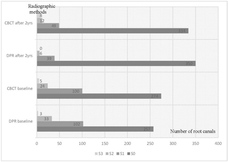
Background and Objectives: There is limited information regarding comparison of long-term dynamics of periapical bone destruction estimated by digital periapical radiography (DPR) and by cone-beam computed tomography (CBCT). This study aimed to compare the radiographically assessed periapical changes of endodontically treated teeth over 2 years of follow-up and to analyse disagreements in periapical lesion size estimates around the same roots using DPR and CBCT. Materials and Methods: A total of 176 endodontically treated teeth, of 128 patients with apical periodontitis, were assessed by DPR and CBCT, at baseline and after 2 years. All periapical radiolucencies were categorised by severity (S0, S1, S2, S3) concerning their size. Descriptive statistics were used to report distribution of the radiolucencies at baseline and at follow-up, and their size transitions over 2 years. Site-specific comparison of the radiolucencies identified by two methods was performed using Z test and Pearson's chi-square test. Results: majority of the detected radiolucencies were scored as S0: 65% and 68% at baseline; 89% and 83% at follow-up, by DPR and CBCT, respectively. Site-specific score comparison showed that disagreements comprised 18% and 20% of the total number of radiolucencies detected by DPR and CBCT, respectively. There were more disagreements between DPR and CBCT within categories S1 and S2 + S3 compared to S0: at baseline, they comprised 17-33% and after two years 62-95% of all detected radiolucencies within the category. 65% of non-matching score transitions over two years occurred between S0 and S1. The CBCT-based evaluation resulted in negative treatment outcomes for 10 more root canals than the DPR-based result. Conclusions: Most remarkable disagreement between DPR and CBCT recordings was observed within the radiolucency categories S2 and S3. However, the diagnostic accuracy of both radiographic methods was questionable as it resulted in a high proportion of non-matching S0-S1 lesion transitions over 2 years.
Keywords: apical periodontitis; cone-beam computed tomography; digital periapical radiography; root canal treatment outcome.
Conflict of interest statement
The authors declare no conflict of interest.
Figures
Similar articles
-
The detection of periapical pathoses using digital periapical radiography and cone beam computed tomography in endodontically retreated teeth - part 2: a 1 year post-treatment follow-up.Int Endod J. 2016 Jul;49(7):623-35. doi: 10.1111/iej.12500. Epub 2015 Jul 29. Int Endod J. 2016. PMID: 26174351
-
The influence of cone-beam computed tomography and periapical radiographic evaluation on the assessment of periapical bone destruction in dog's teeth.Oral Surg Oral Med Oral Pathol Oral Radiol Endod. 2011 Aug;112(2):272-9. doi: 10.1016/j.tripleo.2011.01.031. Epub 2011 May 6. Oral Surg Oral Med Oral Pathol Oral Radiol Endod. 2011. PMID: 21530334
-
The detection of periapical pathosis using digital periapical radiography and cone beam computed tomography - part 2: a 1-year post-treatment follow-up.Int Endod J. 2012 Aug;45(8):711-23. doi: 10.1111/j.1365-2591.2012.02076.x. Int Endod J. 2012. PMID: 22775142
-
Endodontic Periapical Lesion: An Overview on the Etiology, Diagnosis and Current Treatment Modalities.Eur Endod J. 2020 Jul 14;5(2):54-67. doi: 10.14744/eej.2020.42714. eCollection 2020. Eur Endod J. 2020. PMID: 32766513 Free PMC article. Review.
-
New dimensions in endodontic imaging: Part 2. Cone beam computed tomography.Int Endod J. 2009 Jun;42(6):463-75. doi: 10.1111/j.1365-2591.2008.01531.x. Epub 2009 Mar 2. Int Endod J. 2009. PMID: 19298576 Review.
Cited by
-
The outcomes of nonsurgical root canal treatment and retreatment assessed by CBCT: a systematic review and meta-analysis.Saudi Dent J. 2025 Jun 4;37(4-6):14. doi: 10.1007/s44445-025-00021-2. Saudi Dent J. 2025. PMID: 40464818 Free PMC article. Review.
-
Radiomic Parameters in Periapical Lesions: A CBCT Analysis Evaluating Volumetric Size, Cortical Expansion, Erosion, and Shape.Eur Endod J. 2024 Dec;9(4):394-404. doi: 10.14744/eej.2024.45220. Eur Endod J. 2024. PMID: 39704628 Free PMC article.
References
-
- Leonardi Dutra K., Haas L., Porporatti A.L., Flores-Mir C., Santos J.N., Mezzomo L.A., Correa M., Canto G.D. Diagnostic Accuracy of Cone-beam Computed Tomography and Conventional Radiography on Apical Periodontitis: A Systematic Review and Meta-analysis. J. Endod. 2016;42:356–364. doi: 10.1016/j.joen.2015.12.015. - DOI - PubMed
-
- Kanagasingam S., Lim C.X., Yong C.P., Mannocci F., Patel S. Diagnostic accuracy of periapical radiography and cone beam computed tomography in detecting apical periodontitis using histopathological findings as a reference standard. Int. Endod. J. 2017;50:417–426. doi: 10.1111/iej.12650. - DOI - PubMed
MeSH terms
LinkOut - more resources
Full Text Sources
