. 2022 Sep 30;13(10):1645.
doi: 10.3390/mi13101645.

A High-Throughput and Uniform Amplification Method for Cell Spheroids

Affiliations

A High-Throughput and Uniform Amplification Method for Cell Spheroids

Liyuan Liu et al. Micromachines (Basel). .

Abstract

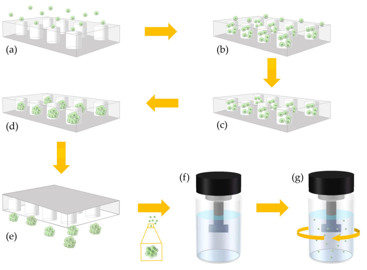
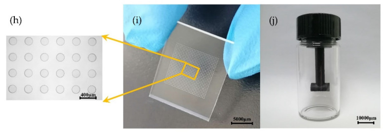
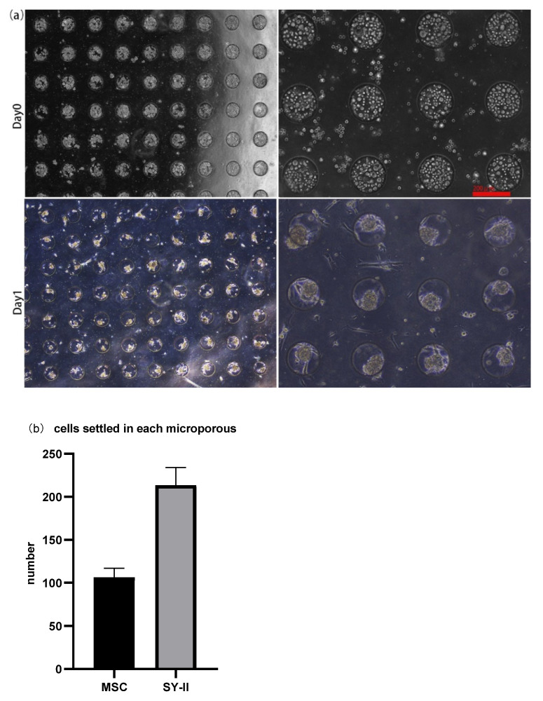
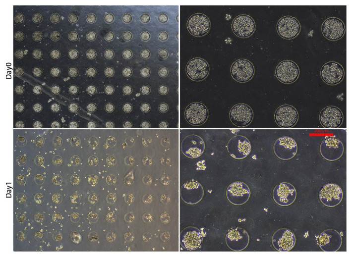
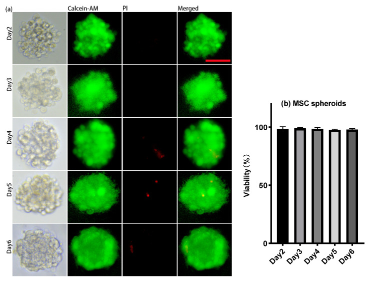
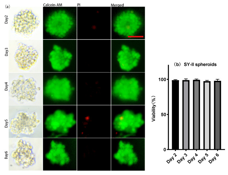
Cell culture is an important life science technology. Compared with the traditional two-dimensional cell culture, three-dimensional cell culture can simulate the natural environment and structure specificity of cell growth in vivo. As such, it has become a research hotspot. The existing three-dimensional cell culture techniques include the hanging drop method, spinner flask method, etc., making it difficult to ensure uniform morphology of the obtained cell spheroids while performing high-throughput. Here, we report a method for amplifying cell spheroids with the advantages of quickly enlarging the culture scale and obtaining cell spheroids with uniform morphology and a survival rate of over 95%. Technically, it is easy to operate and convenient to change substances. These results indicate that this method has the potential to become a promising approach for cell-cell, cell-stroma, cell-organ mutual interaction research, tissue engineering, and anti-cancer drug screening.

Keywords: 3D cell spheroids; cell culture; drug screening; high-throughput; uniform shape.


Conflict of interest statement

The funders had no role in the design of the study; in the collection, analyses, or interpretation of data; in the writing of the manuscript, or in the decision to publish the results.

Figures

Figure 1
Figure 1
Schematic Diagram of High-Throughput and Uniform Cell Sphere Amplification Method. (a) Introducing cells into a microwell chip; (b) cells settle into micropores; (c) washing out the redundant cells on the surface of the microwell chip; (d) the cells in the micropores are aggregated into cell spheres; (e) inverting the microwell chip and taking out the cultured cell spheres; (f) introducing the cell spheres taken out of the microwell chip into a stirring amplification device; (g) dynamically amplifying the cell spheres in a stirring amplification device; (h) microwell structure. D = 200 μm, magnification = 40×, bar = 400 μm; (i) Photographs of the microwell chip, bar = 5000 μm; (j) stirring the amplification device. Bar = 10,000 μm.
Figure 1
Figure 1
Schematic Diagram of High-Throughput and Uniform Cell Sphere Amplification Method. (a) Introducing cells into a microwell chip; (b) cells settle into micropores; (c) washing out the redundant cells on the surface of the microwell chip; (d) the cells in the micropores are aggregated into cell spheres; (e) inverting the microwell chip and taking out the cultured cell spheres; (f) introducing the cell spheres taken out of the microwell chip into a stirring amplification device; (g) dynamically amplifying the cell spheres in a stirring amplification device; (h) microwell structure. D = 200 μm, magnification = 40×, bar = 400 μm; (i) Photographs of the microwell chip, bar = 5000 μm; (j) stirring the amplification device. Bar = 10,000 μm.
Figure 2
Figure 2
(a) The morphology of MSC cells on the day of seeding (Day 0) and spheroid formation (Day 1). On day 0, about 30 μL cell suspension at a concentration of 1 × 10⁷ cells/mL was added to the chip. On day 1, 3D spheroid formation occurred. Magnification = 40× (two pictures on the left); Magnification = 100× (two pictures on the right). Bar = 200 μm. (b) Histograms showed the number of cells that settled into each micropore and eventually formed spheroids of two cell lines, MSC and SYII.
Figure 3
Figure 3
Morphology of SY-II cells on the day of seeding (Day 0) and spheroid formation (Day 1). On day 0, about 30 μL cell suspension with a concentration of 1 × 10⁷ cells/mL was added onto the chip with a pipette. On day 1, 3D spheroid formation occurred. Magnification = 40× (two pictures on the left). Magnification = 100× (two pictures on the right). Bar = 200 μm.
Figure 4
Figure 4
(a) Cell viability test of MSC spheroids: living cells stained with Calcein AM (green) and dead cells stained with PI (red). (b) Histograms showed the cell viability of MSC spheroids on day 2, day 3, day 4, day 5, and day 6. Magnification = 200×, Bar = 50 μm.
Figure 5
Figure 5
(a) Cell viability test of SY-II spheroids: living cells stained with Calcein AM (green) and dead cells stained with PI (red). (b) Histograms showed the cell viability of SY-II spheroids on day 2, day 3, day 4, day 5, and day 6. Magnification = 200×, Bar = 50 μm.

Similar articles

References

    1. Neftel C., Laffy J., Filbin M.G., Hara T., Shore M.E., Rahme G.J., Richman A.R., Silverbush D., Shaw M.L., Hebert C.M., et al. An Integrative Model of Cellular States, Plasticity, and Genetics for Glioblastoma. Cell. 2019;178:835–849.e21. doi: 10.1016/j.cell.2019.06.024. - DOI - PMC - PubMed
    1. Ito M., Komai K., Mise-Omata S., Iizuka-Koga M., Noguchi Y., Kondo T., Sakai R., Matsuo K., Nakayama T., Yoshie O., et al. Brain Regulatory T Cells Suppress Astrogliosis and Potentiate Neurological Recovery. Nature. 2019;565:246–250. doi: 10.1038/s41586-018-0824-5. - DOI - PubMed
    1. Muri J., Heer S., Matsushita M., Pohlmeier L., Tortola L., Fuhrer T., Conrad M., Zamboni N., Kisielow J., Kopf M. The Thioredoxin-1 System Is Essential for Fueling DNA Synthesis during T-Cell Metabolic Reprogramming and Proliferation. Nat. Commun. 2018;9:1851. doi: 10.1038/s41467-018-04274-w. - DOI - PMC - PubMed
    1. Shi Y., Wang Y., Li Q., Liu K., Hou J., Shao C., Wang Y. Immunoregulatory Mechanisms of Mesenchymal Stem and Stromal Cells in Inflammatory Diseases. Nat. Rev. Nephrol. 2018;14:493–507. doi: 10.1038/s41581-018-0023-5. - DOI - PubMed
    1. Fu X., Liu G., Halim A., Ju Y., Luo Q., Song G. Mesenchymal Stem Cell Migration and Tissue Repair. Cells. 2019;8:784. doi: 10.3390/cells8080784. - DOI - PMC - PubMed