Review
. 2022 Oct 13;10(10):2022.
doi: 10.3390/microorganisms10102022.

Schistosomiasis Mansoni-Recruited Eosinophils: An Overview in the Granuloma Context

Affiliations
Review

Schistosomiasis Mansoni-Recruited Eosinophils: An Overview in the Granuloma Context

Kássia K Malta et al. Microorganisms. .

Abstract

Eosinophils are remarkably recruited during schistosomiasis mansoni, one of the most common parasitic diseases worldwide. These cells actively migrate and accumulate at sites of granulomatous inflammation termed granulomas, the main pathological feature of this disease. Eosinophils colonize granulomas as a robust cell population and establish complex interactions with other immune cells and with the granuloma microenvironment. Eosinophils are the most abundant cells in granulomas induced by Schistosoma mansoni infection, but their functions during this disease remain unclear and even controversial. Here, we explore the current information on eosinophils as components of Schistosoma mansoni granulomas in both humans and natural and experimental models and their potential significance as central cells triggered by this infection.

Keywords: Schistosoma mansoni; eosinophil degranulation; eosinophils; granuloma; hepatic granuloma; histopathology; inflammation; schistosomiasis.


Conflict of interest statement

The authors declare no conflict of interest.

Figures
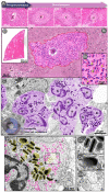
Figure 1
Figure 1
Eosinophils in a hepatic Schistosoma mansoni granuloma. (A) Evolutional stages of the hepatic granuloma and their features: Pre-granulomatous exudative (PE), with an initial infiltration of inflammatory cells in the organization process around the parasite egg (E); Necrotic-exudative (NE), with an area of necrosis in the periovular region around the egg (E) and numerous inflammatory cell types irregularly distributed on subsequent layers; Exudative-productive (EP), more organized and circumferential, with a rich structure of collagen and inflammatory cells concentrated in the periphery; and Productive (P), with a thick band of collagen fibers surrounding the egg (E) and a reduced number of inflammatory cells. (B) A representative whole-slide image of the hepatic tissue showing many granulomas (box). (Bi) A PE granuloma (outlined in red) forms around the parasite egg (E). (Bii) At a higher magnification, observe the accumulation of eosinophils (arrowheads). (C,D) Infiltrated eosinophils (colored in purple in C), seen under transmission electron microscopy (TEM), show their typical ultrastructure with a polylobed nucleus (N) and a high number of cytoplasmic-specific (secretory) granules (colored in yellow in D). Eosinophils represent 60% of all granuloma cells in hepatic NE granulomas [36]. (D) A representative eosinophil shows morphological signs of piecemeal degranulation (PMD), characterized by the presence of enlarged, non-fused granules releasing their content and a high number of transport vesicles (pink), predominantly around emptying granules (yellow). (Di) Intact granules with their unique morphology: a central well-defined electron-dense crystalline core and an outer electron-lucent matrix; (Dii,Diii) Swollen granules with disarranged cores and matrices denote PMD. Liver fragments from experimentally infected (acute phase) mice were prepared for light microscopy (A,B) and conventional TEM (C,D), as before [36]. Histological sections (A,B,Bi,Bii) were stained with Hematoxylin-Eosin. Panels (DDiii) were republished from ref. [36] under the terms of the Creative Commons Attribution License 4.0 (CC-By).
Figure 2
Figure 2
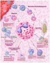
Eosinophil dynamics within a hepatic Schistosoma mansoni granuloma. Mature and immature eosinophils are recruited and migrate from the peripheral blood to target tissues, such as the liver. Undifferentiated eosinophils complete their maturation at the granuloma periphery (extramedullary hematopoietic sites). Eosinophils populate the granuloma and potentially interact with both the egg and other cell populations. Eosinophils may have the following possible functional roles and interactions within the granuloma: (i) destruction of the parasite egg through the secretion of cationic proteins [103]; (ii) interaction with neutrophils and mast cells with a resulting increase in the eosinophil toxicity against the parasite egg [94,95]; (iii) plasma cell sustaining; (iv) interaction with monocytes resulting in decreased monocyte responses [97]; (v) induction of hepatocyte proliferation/liver regeneration; (vi) interaction with macrophages and fibroblasts/myofibroblasts, with an impact on the extracellular matrix (ECM) remodeling and fibrosis induction [6,98]; (vii) enhancement of the immune response as a consequence of the eosinophil Th2 cytokines arsenal [90]; and (viii) interaction with lymphocytes, macrophages, and fibroblasts, which favors eosinophil recruitment, distribution, and activation [89,91].

References

    1. Gryseels B., Polman K., Clerinx J., Kestens L. Human schistosomiasis. Lancet. 2006;368:1106–1118. doi: 10.1016/S0140-6736(06)69440-3. - DOI - PubMed
    1. McManus D.P., Dunne D.W., Sacko M., Utzinger J., Vennervald B.J., Zhou X.N. Schistosomiasis. Nat. Rev. Dis. Prim. 2018;4:13. doi: 10.1038/s41572-018-0013-8. - DOI - PubMed
    1. Butterworth A.E. Immunological aspects of human schistosomiasis. Brit. Med. Bull. 1998;54:357–368. doi: 10.1093/oxfordjournals.bmb.a011693. - DOI - PubMed
    1. Sher A., Coffman R.L., Hieny S., Scott P., Cheever A.W. Interleukin 5 is required for the blood and tissue eosinophilia but not granuloma formation induced by infection with Schistosoma mansoni. Proc. Natl. Acad. Sci. USA. 1990;87:61–65. doi: 10.1073/pnas.87.1.61. - DOI - PMC - PubMed
    1. Gazzinelli G., Lambertucci J.R., Katz N., Rocha R.S., Lima M.S., Colley D.G. Immune responses during human Schistosomiasis mansoni. XI. Immunologic status of patients with acute infections and after treatment. J. Immunol. 1985;135:2121–2127. - PubMed

LinkOut - more resources