Effect of Currently Available Nanoparticle Synthesis Routes on Their Biocompatibility with Fibroblast Cell Lines
- PMID: 36296564
- PMCID: PMC9612073
- DOI: 10.3390/molecules27206972
Effect of Currently Available Nanoparticle Synthesis Routes on Their Biocompatibility with Fibroblast Cell Lines
Erratum in
-
Correction: Mansoor et al. Effect of Currently Available Nanoparticle Synthesis Routes on Their Biocompatibility with Fibroblast Cell Lines. Molecules 2022, 27, 6972.Molecules. 2023 May 18;28(10):4173. doi: 10.3390/molecules28104173. Molecules. 2023. PMID: 37241996 Free PMC article.
Abstract
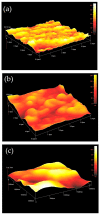
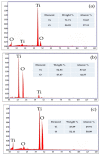
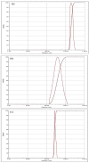
Nanotechnology has acquired significance in dental applications, but its safety regarding human health is still questionable due to the chemicals utilized during various synthesis procedures. Titanium nanoparticles were produced by three novel routes, including Bacillus subtilis, Cassia fistula and hydrothermal heating, and then characterized for shape, phase state, size, surface roughness, elemental composition, texture and morphology by SEM, TEM, XRD, AFM, DRS, DLS and FTIR. These novel titanium nanoparticles were tested for cytotoxicity through the MTT assay. L929 mouse fibroblast cells were used to test the cytotoxicity of the prepared titanium nanoparticles. Cell suspension of 10% DMEM with 1 × 10⁴ cells was seeded in a 96-well plate and incubated. Titanium nanoparticles were used in a 1 mg/mL concentration. Control (water) and titanium nanoparticles stock solutions were prepared with 28 microliters of MTT dye and poured into each well, incubated at 37 °C for 2 h. Readings were recorded on day 1, day 15, day 31, day 41 and day 51. The results concluded that titanium nanoparticles produced by Bacillus subtilis remained non-cytotoxic because cell viability was >90%. Titanium nanoparticles produced by Cassia fistula revealed mild cytotoxicity on day 1, day 15 and day 31 because cell viability was 60−90%, while moderate cytotoxicity was found at day 41 and day 51, as cell viability was 30−60%. Titanium nanoparticles produced by hydrothermal heating depicted mild cytotoxicity on day 1 and day 15; moderate cytotoxicity on day 31; and severe cytotoxicity on day 41 and day 51 because cell viability was less than 30% (p < 0.001). The current study concluded that novel titanium nanoparticles prepared by Bacillus subtilis were the safest, more sustainable and most biocompatible for future restorative nano-dentistry purposes.
Keywords: Bacillus subtilis; Cassia fistula; TiO2; cytotoxicity; nanoparticles.
Conflict of interest statement
The authors declare no conflict of interest.
Figures


Similar articles
-
Synthesis and Characterization of Titanium Oxide Nanoparticles with a Novel Biogenic Process for Dental Application.Nanomaterials (Basel). 2022 Mar 25;12(7):1078. doi: 10.3390/nano12071078. Nanomaterials (Basel). 2022. PMID: 35407196 Free PMC article.
-
Titanium Dioxide Nanoparticle (TiO2 NP) Induces Toxic Effects on LA-9 Mouse Fibroblast Cell Line.Cell Physiol Biochem. 2023 Mar 22;57(2):63-81. doi: 10.33594/000000616. Cell Physiol Biochem. 2023. PMID: 36945889
-
Gellan gum incorporating titanium dioxide nanoparticles biofilm as wound dressing: Physicochemical, mechanical, antibacterial properties and wound healing studies.Mater Sci Eng C Mater Biol Appl. 2019 Oct;103:109770. doi: 10.1016/j.msec.2019.109770. Epub 2019 May 18. Mater Sci Eng C Mater Biol Appl. 2019. PMID: 31349525
-
Functionalized Titanium Nanoparticles Induce Oxidative Stress and Cell Death in Human Skin Cells.Int J Nanomedicine. 2022 Mar 30;17:1495-1509. doi: 10.2147/IJN.S325767. eCollection 2022. Int J Nanomedicine. 2022. PMID: 35388270 Free PMC article.
-
TiO2 nanoparticles co-doped with silver and nitrogen for antibacterial application.J Nanosci Nanotechnol. 2010 Aug;10(8):4868-74. doi: 10.1166/jnn.2010.2225. J Nanosci Nanotechnol. 2010. PMID: 21125821
Cited by
-
Silver Nanoparticles in Dental Applications: A Descriptive Review.Bioengineering (Basel). 2023 Mar 5;10(3):327. doi: 10.3390/bioengineering10030327. Bioengineering (Basel). 2023. PMID: 36978718 Free PMC article. Review.
-
Synthesis of novel titania nanoparticles using corn silky hair fibres and their role in developing a smart restorative material in dentistry.Comput Struct Biotechnol J. 2025 Jan 17;29:29-40. doi: 10.1016/j.csbj.2025.01.005. eCollection 2025. Comput Struct Biotechnol J. 2025. PMID: 39906909 Free PMC article.
-
Role of the novel aloe vera-based titanium dioxide bleaching gel on the strength and mineral content of the human tooth enamel with respect to age.PeerJ. 2024 Sep 18;12:e17779. doi: 10.7717/peerj.17779. eCollection 2024. PeerJ. 2024. PMID: 39308816 Free PMC article.
-
Novel microbial synthesis of titania nanoparticles using probiotic Bacillus coagulans and its role in enhancing the microhardness of glass ionomer restorative materials.Odontology. 2024 Oct;112(4):1123-1134. doi: 10.1007/s10266-024-00921-5. Epub 2024 Mar 30. Odontology. 2024. PMID: 38554219 Free PMC article.
-
Correction: Mansoor et al. Effect of Currently Available Nanoparticle Synthesis Routes on Their Biocompatibility with Fibroblast Cell Lines. Molecules 2022, 27, 6972.Molecules. 2023 May 18;28(10):4173. doi: 10.3390/molecules28104173. Molecules. 2023. PMID: 37241996 Free PMC article.
References
-
- Zhu X., Vo C., Taylor M., Smith B.R. Non-spherical micro- and nanoparticles in nanomedicine. Mater. Horiz. 2019;6:1094–1121. doi: 10.1039/C8MH01527A. - DOI
-
- Colvin V.L., Schlamp M.C., Alivisatos A.P. Light-emitting diodes made from cadmium selenide nanocrystals and a semiconducting polymer. Nature. 1994;370:354–357. doi: 10.1038/370354a0. - DOI
-
- Long M., Wang J., Zhuang H., Zhang Y., Wu H., Zhang J. Performance and mechanism of standard nano-TiO2 (P-25) in photocatalytic disinfection of food borne microorganisms—Salmonella typhimurium and Listeria monocytogenes. Food Control. 2014;39:68–74. doi: 10.1016/j.foodcont.2013.10.033. - DOI
-
- McCullagh C., Robertson J.M., Bahnemann D.W., Robertson P.K. The application of TiO2 photocatalysis for disinfection of water contaminated with pathogenic micro-organisms: A review. Res. Chem. Intermed. 2007;33:359–375. doi: 10.1163/156856707779238775. - DOI
MeSH terms
Substances
LinkOut - more resources
Full Text Sources
Miscellaneous

