. 2022 Oct 11;12(20):3561.
doi: 10.3390/nano12203561.

Different Strategies to Attenuate the Toxic Effects of Zinc Oxide Nanoparticles on Spermatogonia Cells

Affiliations

Different Strategies to Attenuate the Toxic Effects of Zinc Oxide Nanoparticles on Spermatogonia Cells

Mariana Vassal et al. Nanomaterials (Basel). .

Abstract

Zinc oxide nanoparticles (ZnO NPs) are one of the most used nanoparticles due to their unique physicochemical and biological properties. There is, however, a growing concern about their negative impact on male reproductive health. Therefore, in the present study, two different strategies were used to evaluate the recovery ability of spermatogonia cells from the first stage of spermatogenesis (GC-1 spg cell line) after being exposed to a cytotoxic concentration of ZnO NPs (20 µg/mL) for two different short time periods, 6 and 12 h. The first strategy was to let the GC-1 cells recover after ZnO NPs exposure in a ZnO NPs-free medium for 4 days. At this phase, cell viability assays were performed to evaluate whether this period was long enough to allow for cell recovery. Exposure to ZnO NPs for 6 h and 12 h induced a decrease in viability of 25% and 41%, respectively. However, the recovery period allowed for an increase in cell viability from 16% to 25% to values as high as 91% and 84%. These results strongly suggest that GC-1 cells recover, but not completely, given that the cell viability does not reach 100%. Additionally, the impact of a synthetic chalcone (E)-3-(2,6-dichlorophenyl)-1-(2-hydroxyphenyl)prop-2-en-1-one (1) to counteract the reproductive toxicity of ZnO NPs was investigated. Different concentrations of chalcone 1 (0-12.5 µM) were used before and during exposure of GC-1 cells to ZnO NPs to mitigate the damage induced by NPs. The protective ability of this compound was evaluated through viability assays, levels of DNA damage, and cytoskeleton dynamics (evaluating the acetylated α-tubulin and β-actin protein levels). The results indicated that the tested concentrations of chalcone 1 can attenuate the genotoxicity induced by ZnO NPs for shorter exposure periods (6 h). Chalcone 1 supplementation also increased cell viability and stabilized the microtubules. However, the antioxidant potential of this compound remains to be elucidated. In conclusion, this work addressed the main cytotoxic effects of ZnO NPs on a spermatogonia cell line and analyzed two different strategies to mitigate this damage, which represent a significant contribution to the field of male fertility.

Keywords: DNA damage; antioxidant; chalcone; cytoskeleton; cytotoxicity; male infertility; oxidative stress; reversibility; zinc oxide nanoparticles.


Conflict of interest statement

The authors declare no conflict of interest.

Figures

Figure 1
Figure 1
Structure of the synthesized chalcone 1 [(E)-3-(2,6-dichlorophenyl)-1-(2-hydroxyphenyl)prop-2-en-1-one]. The 1,3-diarylprop-2-en-1-one fragment is marked in red.
Figure 2
Figure 2
Particle size distribution and SEM images of the ZnO powder used.
Figure 3
Figure 3
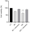
GC-1 cells recovery upon NPs exposure. Results from viability assay analysis of GC-1 cells after their incubation with 20 µg/mL of ZnO NPs, for 6 and 12 h, and after a recovery period of 4 days. The percentage of viable cells for each condition was plotted as the mean ± SEM of four independent experiments. Values are expressed as arbitrary units, and the cell viability of the control condition was given a value of 100. ** p < 0.01 compared to the control group, using one way ANOVA. CT: control.
Figure 4
Figure 4
Cell viability assay of cells treated with chalcone 1 (for the sake of simplicity, the chalcone is indicated in the graphics using only the number 1). Changes in the viability of the GC-1 cells after being treated with different concentrations of chalcone 1 (0–25 µM) for (A) 7 h and (B) 13 h. The percentage of viable cells for each condition was plotted as mean ± SEM of five independent experiments, performed in duplicate. Values are expressed as arbitrary units, and the cell viability of the control condition is given a value of 100. ** p < 0.01 compared to the control group, using a nonparametric Kruskal–Wallis one-way ANOVA test.
Figure 5
Figure 5
Cell viability assay of cells co-exposed with chalcone 1 and ZnO NPs. Changes in the viability of the GC-1 cells treated with 20 µg/mL of ZnO NPs in the absence and presence of different concentrations of chalcone 1 (0–12.5 µM) after (A) 6 and (B) 12 h of exposure. The percentage of viable cells for each condition was plotted as mean ± SEM of five independent experiments, performed in duplicate. Values are expressed as arbitrary units, and the cell viability of the control condition is given a value of 100. **** p < 0.0001 compared to the control group, # p < 0.05 and ### p < 0.001 compared to the group treated with ZnO NPs, using one-way ANOVA. NPs: nanoparticles.
Figure 6
Figure 6
Immunoblotting analysis of γ-H2AX (Ser139) intracellular levels. This assay allows for the assessment of the DNA damage after the incubation of GC-1 cells with 20 µg/mL of ZnO NPs and different concentrations of chalcone 1 (0–12.5 µM) per se, and the protective effects of chalcone 1 against the genotoxicity induced by the NPs after (A) 6 h and (B) 12 h of co-exposure with ZnO NPs. The intracellular protein levels of γ-H2AX were estimated in relation to protein levels detected in the control condition. Ponceau S staining was used to assess gel loading. * p < 0.05, *** p < 0.001, and **** p < 0.0001 compared to the control group, using one-way ANOVA. All data were expressed as the mean ± SEM of four independent experiments. NPs: nanoparticles.
Figure 7
Figure 7
Immunoblotting analysis of cytoskeleton proteins. Acetylated α-tubulin intracellular protein levels of GC-1 cells treated with ZnO NPs (20 µg/mL) and chalcone 1 (0–12.5 µM) for 6 (A) and 12 h (B). The intracellular protein levels of acetylated α-tubulin were estimated in relation to protein levels detected in the control condition. Ponceau S staining was used to assess gel loading. All data were expressed as the mean ± SEM of four independent experiments. ** p < 0.01, *** p < 0.001 and, **** p < 0.0001 compared to the control group, using one-way ANOVA, and ## p < 0.01 compared to the group treated with ZnO NPs using the Student’s t-test. β-actin intracellular protein levels of GC-1 cells treated with ZnO NPs (20 µg/mL) and chalcone 1 (0–12.5 µM) for 6 (C) and 12 h (D). The intracellular protein levels of β-actin were estimated in relation to protein levels detected in the control condition. Ponceau S staining was used to assess gel loading. All data were expressed as mean ± SEM of three independent experiments. No significant differences were detected between groups when analyzed by one-way ANOVA. NPs: nanoparticles.

References

    1. Pinho A.R., Martins F., Costa M.E.V., Senos A.M.R., Silva O.A.B.D.C.E., Pereira M.D.L., Rebelo S. In Vitro Cytotoxicity Effects of Zinc Oxide Nanoparticles on Spermatogonia Cells. Cells. 2020;9:1081. doi: 10.3390/cells9051081. - DOI - PMC - PubMed
    1. Barkhoradi A., Hekmatimoghaddam S., Jebali A., Khalili M., Talebi A., Noorani M. Effect of Zinc Oxide Nanoparticles on Viability of Human Spermatozoa. Iran J. Reprod. Med. 2013;11:767–771. - PMC - PubMed
    1. Shen J., Yang D., Zhou X., Wang Y., Tang S., Yin H., Wang J., Chen R., Chen J. Role of Autophagy in Zinc Oxide Nanoparticles-Induced Apoptosis of Mouse LEYDIG Cells. Int. J. Mol. Sci. 2019;20:4042. doi: 10.3390/ijms20164042. - DOI - PMC - PubMed
    1. Kalpana V.N., Rajeswari V.D. A Review on Green Synthesis, Biomedical Applications, and Toxicity Studies of ZnO NPs. Bioinorg. Chem. Appl. 2018;2018:3569758. doi: 10.1155/2018/3569758. - DOI - PMC - PubMed
    1. Fontecha-Umaña F., Ríos-Castillo A.G., Ripolles-Avila C., Rodríguez-Jerez J.J. Antimicrobial Activity and Prevention of Bacterial Biofilm Formation of Silver and Zinc Oxide Nanoparticle-Containing Polyester Surfaces at Various Concentrations for Use. Foods. 2020;9:442. doi: 10.3390/foods9040442. - DOI - PMC - PubMed