. 2022 Sep 27;15(10):1191.
doi: 10.3390/ph15101191.

Pretargeted Imaging beyond the Blood-Brain Barrier-Utopia or Feasible?

Affiliations

Pretargeted Imaging beyond the Blood-Brain Barrier-Utopia or Feasible?

Sara Lopes van den Broek et al. Pharmaceuticals (Basel). .

Abstract

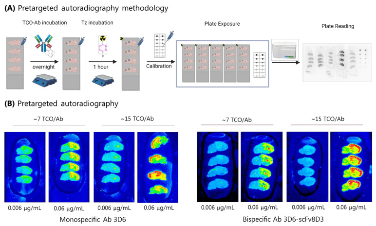
Pretargeting is a promising nuclear imaging technique that allows for the usage of antibodies (Abs) with enhanced imaging contrast and reduced patient radiation burden. It is based on bioorthogonal chemistry with the tetrazine ligation-a reaction between trans-cyclooctenes (TCOs) and tetrazines (Tzs)-currently being the most popular reaction due to its high selectivity and reactivity. As Abs can be designed to bind specifically to currently 'undruggable' targets such as protein isoforms or oligomers, which play a crucial role in neurodegenerative diseases, pretargeted imaging beyond the BBB is highly sought after, but has not been achieved yet. A challenge in this respect is that large molecules such as Abs show poor brain uptake. Uptake can be increased by receptor mediated transcytosis; however, it is largely unknown if the achieved brain concentrations are sufficient for pretargeted imaging. In this study, we investigated whether the required concentrations are feasible to reach. As a model Ab, we used the bispecific anti-amyloid beta (Aβ) anti-transferrin receptor (TfR) Ab 3D6scFv8D3 and conjugated it to a different amount of TCOs per Ab and tested different concentrations in vitro. With this model in hand, we estimated the minimum required TCO concentration to achieve a suitable contrast between the high and low binding regions. The estimation was carried out using pretargeted autoradiography on brain sections of an Alzheimer's disease mouse model. Biodistribution studies in wild-type (WT) mice were used to correlate how different TCO/Ab ratios alter the brain uptake. Pretargeted autoradiography showed that increasing the number of TCOs as well as increasing the TCO-Ab concentration increased the imaging contrast. A minimum brain concentration of TCOs for pretargeting purposes was determined to be 10.7 pmol/g in vitro. Biodistribution studies in WT mice showed a brain uptake of 1.1% ID/g using TCO-3D6scFv8D3 with 6.8 TCO/Ab. According to our estimations using the optimal parameters, pretargeted imaging beyond the BBB is not a utopia. Necessary brain TCO concentrations can be reached and are in the same order of magnitude as required to achieve sufficient contrast. This work gives a first estimate that pretargeted imaging is indeed possible with antibodies. This could allow the imaging of currently 'undruggable' targets and therefore be crucial to monitor (e.g., therapies for intractable neurodegenerative diseases).

Keywords: CNS; PET; antibody; brain; pretargeting; trans-cyclooctene.


Conflict of interest statement

The authors declare no conflict of interest.

Figures

Figure 1
Figure 1
(A) An illustration of receptor-mediated transcytosis (RTM). A bispecific ligand binds to its carrier protein and is transported into the brain by transcytosis. (B) A schematic overview of pretargeted imaging beyond the BBB. The TCO-Ab is injected first and accumulated at its target within the brain. In a second step, a radiolabeled Tz is administered. This Tz crosses the BBB and reacts with the TCO-Ab. This ligation results in a radiolabeled Ab that can be imaged. (C) An overview of the factors contributing to pretargeting in the brain. Previous work describes the identification of animal models and BBB-permeable Tzs. In this work, parameters regarding BBB-permeable TCO-Abs were investigated.
Figure 2
Figure 2
(A) SDS-PAGE with 111In-Tz and TCO-Abs to quantify the TCO load. Activity at the bottom corresponds to the 111In-Tz and the activity bands at the top to the radiolabeled Ab. The difference in radioactivity was used for the TCO quantification (Supplementary Materials). The lanes on the left correspond to ~6 TCO/Ab, and the middle lanes to ~18 and ~19 TCO/Ab TCO/Ab. The lane on the right is the 111In-Tz without Ab (control). (B) Different TCO-loading can be reached using different equivalents of TCO. The saturation of TCO-loading was reached around 20 TCOs. (C) ELISA for Aβ with Ab 3D6 and TCO-3D6scFv8D3. Affinity was reduced with an increased amount of TCOs.
Figure 3
Figure 3
(A) The pretargeted autoradiography workflow. TCO-Abs was applied first and incubated overnight. Then, the radiolabeled Tz was applied. Slides were washed and a calibration curve was added before slide exposure to the phosphor plates. Phosphor plates were read-out to provide the autoradiography images. (B) Examples of the autoradiography results with TCO-3D6 and TCO-3D5scFv8D3. Increasing TCO-loading and TCO-Ab concentration increased the imaging contrast. Similar results were observed for TCO-3D6 and TCO-3D5scFv8D3.
Figure 4
Figure 4
(A) Experimental setup biodistribution study. TCO-3D6scFv8D3 was reacted in vitro to 18F-tetrazine to obtain the ligated product. 18F-3D6scFv8D3 was injected into the WT mice. After two hours, the mice were euthanized, and the organs were collected and measured by gamma counting. (B) Brain uptake measured by gamma counting. RH = Right hemisphere. LH = Left hemisphere (C) Biodistribution of 18F-3D6scFv8D3. (D) pH stability of TCO-3D6scFv8D3 showing a stability at over three days at pH 5, 6, and 7.4. TCO-3D6scFv8D3 was incubated with an excess of 111In-Tz and the TCO-reactivity was monitored by evaluating Tz ligation using radio-HPLC.

References

    1. Khalil M.M., Tremoleda J.L., Bayomy T.B., Gsell W. Molecular SPECT Imaging: An Overview. Int. J. Mol. Imaging. 2011;2011:796025. doi: 10.1155/2011/796025. - DOI - PMC - PubMed
    1. Basu S., Kwee T.C., Surti S., Akin E.A., Yoo D., Alavi A. Fundamentals of PET and PET/CT imaging. Ann. N. Y. Acad. Sci. 2011;1228:1–18. doi: 10.1111/j.1749-6632.2011.06077.x. - DOI - PubMed
    1. Piel M., Vernaleken I., Rösch F. Positron emission tomography in CNS drug discovery and drug monitoring. J. Med. Chem. 2014;57:9232–9258. doi: 10.1021/jm5001858. - DOI - PubMed
    1. Ametamey S.M., Honer M., Schubiger P.A. Molecular imaging with PET. Chem. Rev. 2008;108:1501–1516. doi: 10.1021/cr0782426. - DOI - PubMed
    1. Theek B., Rizzo L.Y., Ehling J., Kiessling F., Lammers T. The theranostic path to personalized nanomedicine. Clin. Transl. Imaging. 2014;2:67–76. doi: 10.1007/s40336-014-0051-5. - DOI - PMC - PubMed