Review
. 2022 Oct 19;15(10):1283.
doi: 10.3390/ph15101283.

Strategies for Improving Peptide Stability and Delivery

Affiliations
Review

Strategies for Improving Peptide Stability and Delivery

Othman Al Musaimi et al. Pharmaceuticals (Basel). .

Abstract

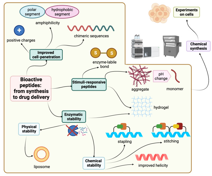
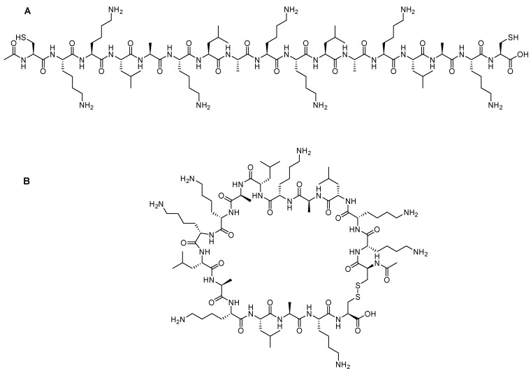
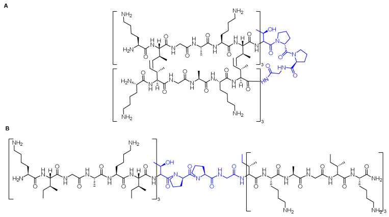
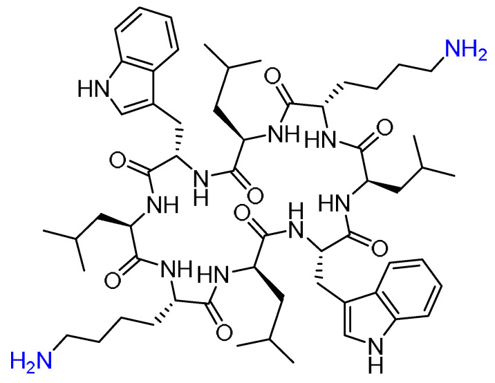
Peptides play an important role in many fields, including immunology, medical diagnostics, and drug discovery, due to their high specificity and positive safety profile. However, for their delivery as active pharmaceutical ingredients, delivery vectors, or diagnostic imaging molecules, they suffer from two serious shortcomings: their poor metabolic stability and short half-life. Major research efforts are being invested to tackle those drawbacks, where structural modifications and novel delivery tactics have been developed to boost their ability to reach their targets as fully functional species. The benefit of selected technologies for enhancing the resistance of peptides against enzymatic degradation pathways and maximizing their therapeutic impact are also reviewed. Special note of cell-penetrating peptides as delivery vectors, as well as stapled modified peptides, which have demonstrated superior stability from their parent peptides, are reported.

Keywords: cell-penetrating peptides; hydrogel; peptides; self-assembled peptides; stapled peptides; stitched peptides.


Conflict of interest statement

The authors declare no conflict of interest.

Figures

Figure 1
Figure 1
Strategies and tactics to improve peptide stability and delivery. Created with Biorender.com.
Figure 2
Figure 2
TAT sequence showing the tentative functional regions (adopted from [26]).
Figure 3
Figure 3
Different sequences. The region that corresponds to the third helix of the homeodomain is shown in blue. The two new F residues that replaced the W are underlined.
Figure 4
Figure 4
Transportan chimeric peptide sequence. Red: the sequence from galanin neuropeptide; Blue the sequence from the wasp venom peptide toxin (mastoparan).
Figure 5
Figure 5
Ruthenium catalyzed olefin metathesis (RCM). (A) O-allyl serine residues by Grubbs [59]. (B) The α,α-disubstitution, olefin tether by Verdine [60].
Figure 6
Figure 6
The sequences of the parent Ascaphin-8 peptide and four analogs with different stapling positions. Fmoc-S5-OH (Fmoc-(S)-2-(4-pentenyl)Ala-OH) and Fmoc-R8-OH (Fmoc-(R)-2-(7-octenyl)Ala-OH) are shown at the right in red and blue, respectively.
Figure 7
Figure 7
The sequences of the parent hCD81-LEL peptide and five analogs with different stapling positions. Fmoc-S5-OH and Fmoc-R8-OH are shown at the right in red and blue, respectively. The parent peptide was identified and synthesized by Dhanasekaran et al. [62]. For S5 and R8, refer to the legend of Figure 6.
Figure 8
Figure 8
T649v parent peptides and the corresponding stapled modified analogs. X in blue represents the stapling positions.
Figure 9
Figure 9
BID-BH3 parent peptide and the corresponding five analogs with different stapling positions. Nor: Norleucine. Fmoc-R5-OH (Fmoc-(R)-2-(4-pentenyl)Ala-OH) and Fmoc-S8-OH (Fmoc-(S)-2-(7-octenyl)Ala-OH) are shown at the right in pink and green, respectively. For S5 refer to the legend of Figure 6.
Figure 10
Figure 10
SAH-p53 parent peptide and ten corresponding analogs with different stapling positions. Both Asn and Gln that replaced Asp and Glu residues are shown in green; Arg that replaced Lys is shown in pink; Lys that replaced Leu is shown in orange. For S5 refer to the legend of Figure 6.
Figure 11
Figure 11
Chemical structure of Gramicidin S (GraS).
Figure 12
Figure 12
Chemical structures of polymyxin B (PMB) and the three amphiphile peptides (PA): threonine (T)-containing PA (TPA), glutamic acid (E)-containing PA (EPA), and beta-sheet-forming PA (BPA).
Figure 13
Figure 13
Chemical structure of (A) Linear KLA-SS peptide (B); Cyclic KLA-SS peptide.
Figure 14
Figure 14
Chemical structure of: (A) ASCP1; (B) ASCP2. The linker is shown in blue.
Figure 15
Figure 15
Chemical structure of the cyclic peptide comprising alternating L and D amino acids. Anchoring points are shown in blue.

References

    1. Al Musaimi O., Al Shaer D., De la Torre B.G., Albericio F. 2017 FDA Peptide Harvest. Pharmaceuticals. 2018;11:42. doi: 10.3390/ph11020042. - DOI - PMC - PubMed
    1. Jensen S.M., Potts G.K., Ready D.B., Patterson M.J. Specific MHC-I Peptides Are Induced Using PROTACs. Front. Immunol. 2018;9:2697. doi: 10.3389/fimmu.2018.02697. - DOI - PMC - PubMed
    1. Jeong W.J., Bu J., Kubiatowicz L.J., Chen S.S., Kim Y., Hong S. Peptide-nanoparticle conjugates: A next generation of diagnostic and therapeutic platforms? Nano Converg. 2018;5:38. doi: 10.1186/s40580-018-0170-1. - DOI - PMC - PubMed
    1. Al Shaer D., Al Musaimi O., Albericio F., de la Torre B.G. 2018 FDA Tides Harvest. Pharmaceuticals. 2019;12:52. doi: 10.3390/ph12020052. - DOI - PMC - PubMed
    1. Du A.W., Stenzel M.H. Drug carriers for the delivery of therapeutic peptides. Biomacromolecules. 2014;15:1097–1114. doi: 10.1021/bm500169p. - DOI - PubMed

LinkOut - more resources