. 2022 Oct 6;14(10):2125.
doi: 10.3390/pharmaceutics14102125.

Continuous NPWT Regulates Fibrosis in Murine Diabetic Wound Healing

Affiliations

Continuous NPWT Regulates Fibrosis in Murine Diabetic Wound Healing

Mengfan Wu et al. Pharmaceutics. .

Abstract

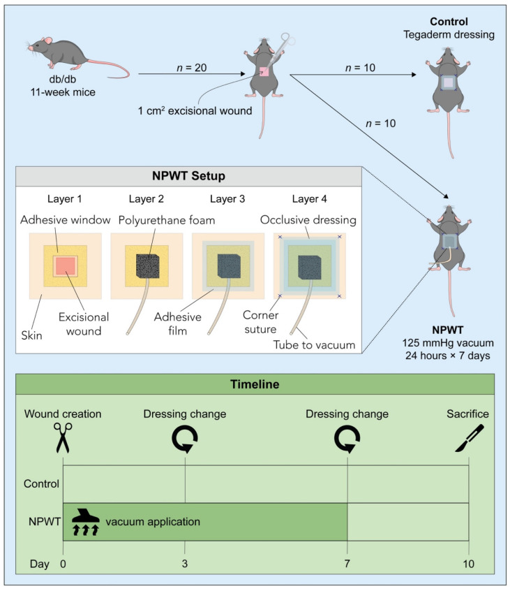
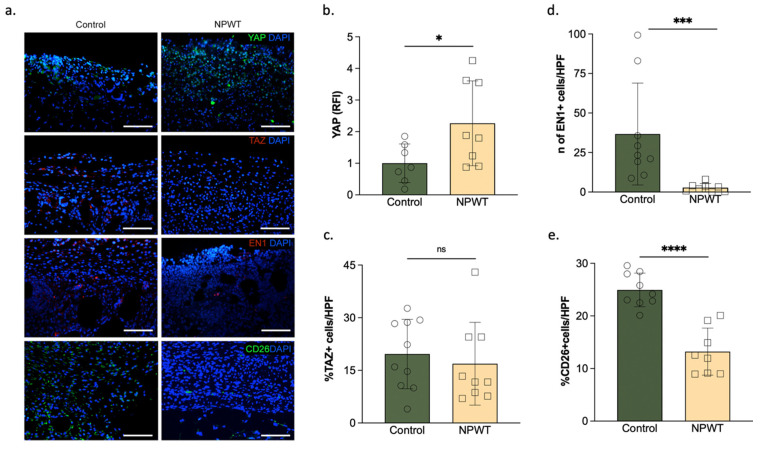
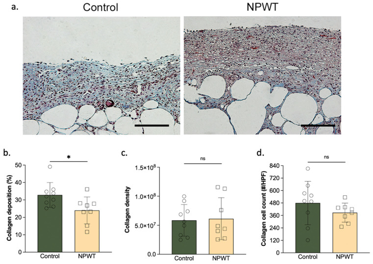
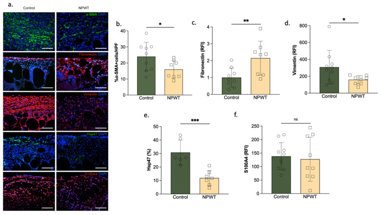
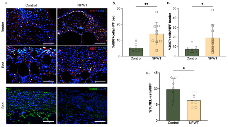
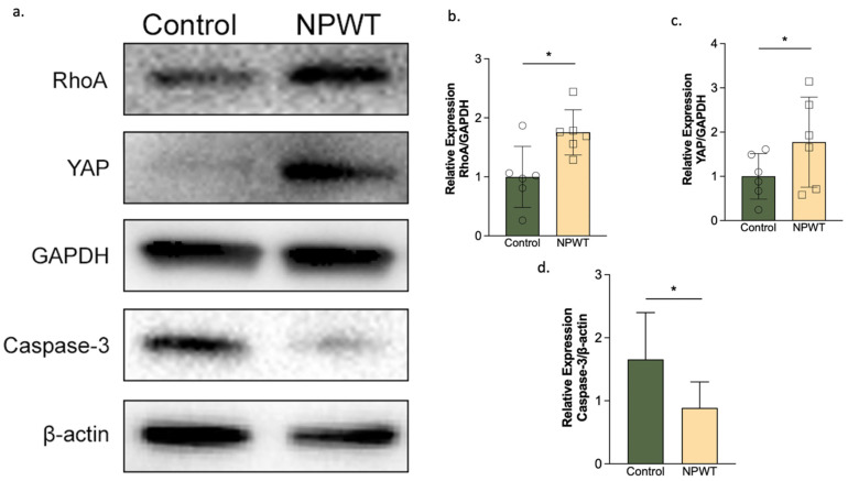
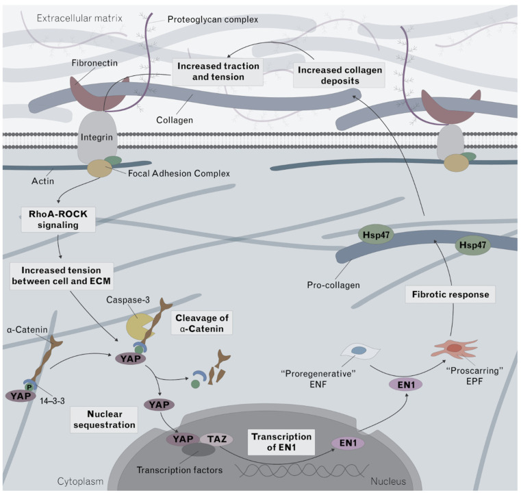
Scarring is associated with significant morbidity. The mechanical signaling factor yes-associated protein (YAP) has been linked to Engrailed-1 (En1)-lineage positive fibroblasts (EPFs), a pro-scarring fibroblast lineage, establishing a connection between mechanotransduction and fibrosis. In this study, we investigate the impact of micromechanical forces exerted through negative pressure wound therapy (NPWT) on the pathophysiology of fibrosis. Full-thickness excisional dorsal skin wounds were created on diabetic (db/db) mice which were treated with occlusive covering (control) or NPWT (continuous, −125 mmHg, 7 days; NPWT). Analysis was performed on tissue harvested 10 days after wounding. NPWT was associated with increased YAP (p = 0.04) but decreased En1 (p = 0.0001) and CD26 (p < 0.0001). The pro-fibrotic factors Vimentin (p = 0.04), α-SMA (p = 0.04) and HSP47 (p = 0.0008) were decreased with NPWT. Fibronectin was higher (p = 0.01) and collagen deposition lower in the NPWT group (p = 0.02). NPWT increased cellular proliferation (p = 0.002) and decreased apoptosis (p = 0.03). Western blotting demonstrated increased YAP (p = 0.02) and RhoA (p = 0.03) and decreased Caspase-3 (p = 0.03) with NPWT. NPWT uncouples YAP from EPF activation, through downregulation of Caspace-3, a pro-apoptotic factor linked to keloid formation. Mechanotransduction decreases multiple pro-fibrotic factors. Through this multifactorial process, NPWT significantly decreases fibrosis and offers promising potential as a mode to improve scar appearance.

Keywords: NPWT; YAP; caspase3; fibrosis; mechanotransduction; scarring; tissue regeneration; wound healing.


Conflict of interest statement

D.P.O. receives research funding through sponsored research agreement to Brigham and Women’s Hospital from KCI (Kinetic Concepts Inc.).

Figures

Figure 1
Figure 1
Study design. 20 db/db mice received a full-thickness dorsal skin excisional wound (1 × 1 cm). 10 mice were treated with NPWT (NPWT group), and 10 mice were covered with an occlusive dressing (control group). The NPWT setup displays the setup of the different layers and the timeline depicts the time points of NPWT application, dressing change and sacrifice.
Figure 2
Figure 2
YAP, TAZ, En1 and CD26 levels. (a) Immunofluorescent staining of the wound bed. anti-YAP, anti-TAZ, anti-En1 and anti-CD26 with DAPI stained sections of the wound bed of the two groups on day 10. Scale bar = 100 µm. (b) Relative fluorescence intensity of YAP. YAP presence significantly differed between the two groups (NPWT: 2.3 ± 1.3 RFI vs. control:1 ± 0.6; p = 0.04). (c) Percentage of TAZ + ve cells. There were no differences in the presence of TAZ between the two groups. (d) Percentage of En1 + ve cells. En1 was significantly lower in the NPWT group (NPWT:2.8 ± 2.6% En1 + cells vs. control:37 ± 32% En1 + cells; p = 0.01). (e) Percentage of CD26 + ve cells. CD26 was significantly lower in the NPWT group (NPWT: 2.8 ± 2.6% CD26 + cells vs. control:37 ± 32% CD26 + cells; p < 0.0001). Number of circles/squares per bar equals sample size (n = 7–10) with each circle/square representing the average of three HPF measurements. Where ns equals p > 0.05, * equals p ≤ 0.05, *** equals p ≤ 0.001 and **** equals p ≤ 0.0001.
Figure 3
Figure 3
Collagen deposition. (a) Representative sections. MT-stained wound beds of the two groups on day 10. Scale bar = 100 µm. (b) Collagen deposition. Collagen deposition significantly differed between the two groups (NPWT: 24 ± 7.7% vs. control: 33 ± 7.1; p = 0.02). (c) Collagen density. There were no significant differences in collagen density between the two groups (NPWT: 6.1 ± 3.6 × 10⁷ vs. control: 5.9 ± 2.7 × 10⁷; p = 0.86). (d) Collagen cell count. There were no significant differences in collagen cell count (NPWT: 387 ± 91 cells/HPF vs. control: 480 ± 208 × 107 cells/HPF; p = 0.26). Number of circles/squares per bar equals sample size (n = 8–9) with each circle/square representing the average of three HPF measurements. Where ns equals p > 0.05 and * equals p ≤ 0.05.
Figure 4
Figure 4
αSMA, Fibronectin, Vimentin, Hsp47 and S100A4 deposition. (a) Representative sections. Fibronectin, Vimentin, Hsp47 and S100A4 -stained wound beds of the two groups on day 10. Scale bar = 100 µm. (b) αSMA deposition. αSMA deposition was significantly higher in the control group (NPWT:16 ± 5% αSMA + cells vs. control: 24 ± 8%αSMA + cells; p = 0.04). (c) Fibronectin deposition. Fibronectin deposition significantly differed between the two groups (NPWT:2.2 ± 1 RFI vs. control: 1 ± 0.6; p = 0.01). (d) Vimentin deposition. Vimentin deposition significantly differed between the two groups (NPWT: 160 ± 50 RFI vs. control: 325 ± 203 RFI; p = 0.04). (e). Hsp47 deposition. Hsp47 deposition significantly differed between the two groups (NPWT:12 ± 5.6% vs. control: 31 ± 9.2%; p = 0.0008). (f) S100A4 deposition. No differences were noted in S100A4 deposition between the two groups (NPWT:127 ± 81 RFI vs. control: 138 ± 50 RFI; p = 0.73). Number of circles/squares per bar indicates sample size (n = 8–9). Each circle/square represents the average of three HPF measurements. Where * equals p ≤ 0.05, ** equals p ≤ 0.01, and *** equals p ≤ 0.001.
Figure 5
Figure 5
Cell turnover. (a) Representative sections. anti-Ki67, Tunel and DAPI stained sections of the wound bed and border of the two groups on day 10. Scale bar = 100 µm. (b) Cellular proliferation of the wound bed. Cellular proliferation of the wound bed significantly differed between the two groups (NPWT: 14 ± 7.2% Ki67 + cells/HPF vs. control: 5.2 ± 2.4% Ki67 + cells; p = 0.002). (c) Cellular proliferation of the wound border. Cellular proliferation of the wound border significantly differed between the two groups (NPWT: 19 ± 14% Ki67 + cells/HPF vs. control:7.3 ± 3.5% Ki67 + cells; p = 0.03). (d) Cellular apoptosis. Cellular apoptosis significantly differed between the two groups (NPWT: 19 ± 6% TUNEL +/HPF vs. control: 29 ± 10% TUNEL +/HPF; p = 0.03). Number of circles/squares per bar indicates sample size (n = 8–10). Each circle/square represents the average of three HPF measurements. Where * equals p ≤ 0.05 and ** equals p ≤ 0.01.
Figure 6
Figure 6
YAP, RhoA and Caspase-3 expression. (a) Representative Western blots. YAP, RhoA and Caspase-3 protein expression in the two groups on day 10. (b) RhoA relative expression. Densitometry of the ratios of RhoA to GAPDH (n = 6; p = 0.03) with levels visualized as bar graphs. The NPWT group had significantly higher RhoA levels than the control group. (c) YAP relative expression. Densitometry of the ratios of YAP to GAPDH (n = 6; p = 0.02) with levels indicated as bar graphs. The NPWT group had significantly higher YAP levels than the control group. (d) Caspase-3 relative expression. Densitometry of the ratios of Caspase-3 to β-Actin (n = 6; p = 0.03) with the levels indicated as bar graphs. Caspase-3 levels were significantly higher in the control compared to the NPWT group. Where * equals p ≤ 0.05.
Figure 7
Figure 7
Proposed mechanism of action of NPWT in fibrosis. The normal fibrotic response that occurs during wound healing, and which becomes upregulated in hypertrophic scarring, is summarized in this schematic. Mechanotransduction, in the form of increased tension between the cell and the extracellular matrix, is hypothesized to lead to an upregulation of YAP. Within the cytoplasm, YAP is bound to a-catenin via 14-3-3 which prevents its nuclear sequestration. Fibrosis-promoting signals upregulated caspase-3 which cleaves a-catenin, and allows YAP to translocate into the nucleus. YAP is then able to promote transcription of En1 which induces a switch of the fibroblast phenotype, from pro-regenerative to pro-fibrotic. The fibrotic response results in an increase in the conversion of pro-collagen to collagen. The increased collagen deposition and increase in scarring then further amplifies the loop. In this study, we demonstrate that although NPWT increased the expression of YAP, En1 was decreased. The decrease in Caspase-3 would help explain this de-coupling, as decreased caspase-3 would result in decreased cleavage of a-catenin, decreased nuclear sequestration of YAP and decreased En1 transcription, as identified in our study. A decreased fibrotic response would result in decreased collagen formation. Hsp47 which is required for pro-collagen to be converted to collagen was also identified as decreased in this study, further supporting the decoupling mechanism. YSP, yes-associated protein; EN1, Engrailed-1; RhoA–ROCK, RhoA and Rho-associated protein kinase.

References

    1. Gurtner G.C., Werner S., Barrandon Y., Longaker M.T. Wound repair and regeneration. Nature. 2008;453:314–321. doi: 10.1038/nature07039. - DOI - PubMed
    1. Walmsley G.G., Maan Z.N., Wong V.W., Duscher D., Hu M.S., Zielins E.R., Wearda T., Muhonen E., McArdle A., Tevlin R.M.B., et al. Scarless wound healing: Chasing the holy grail. Plast. Reconstr. Surg. 2015;135:907–917. doi: 10.1097/PRS.0000000000000972. - DOI - PubMed
    1. Lorenz H.P., Longaker M., Perkocha L., Jennings R., Harrison M., Adzick N. Scarless wound repair: A human fetal skin model. Development. 1992;114:253–259. doi: 10.1242/dev.114.1.253. - DOI - PubMed
    1. Ud-Din S., Volk S.W., Bayat A. Regenerative healing, scar-free healing and scar formation across the species: Current concepts and future perspectives. Exp. Dermatol. 2014;23:615–619. doi: 10.1111/exd.12457. - DOI - PubMed
    1. Armstrong J.R., Ferguson M.W.J. Ontogeny of the skin and the transition from scar-free to scarring phenotype during wound healing in the pouch young of a marsupial, Monodelphis domestica. Dev. Biol. 1995;169:242–260. doi: 10.1006/dbio.1995.1141. - DOI - PubMed