. 2022 Sep 21;10(10):1582.
doi: 10.3390/vaccines10101582.

Nonhuman Primates Are Protected against Marburg Virus Disease by Vaccination with a Vesicular Stomatitis Virus Vector-Based Vaccine Prepared under Conditions to Allow Advancement to Human Clinical Trials

Affiliations

Nonhuman Primates Are Protected against Marburg Virus Disease by Vaccination with a Vesicular Stomatitis Virus Vector-Based Vaccine Prepared under Conditions to Allow Advancement to Human Clinical Trials

Christopher L Cooper et al. Vaccines (Basel). .

Abstract

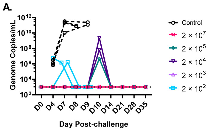
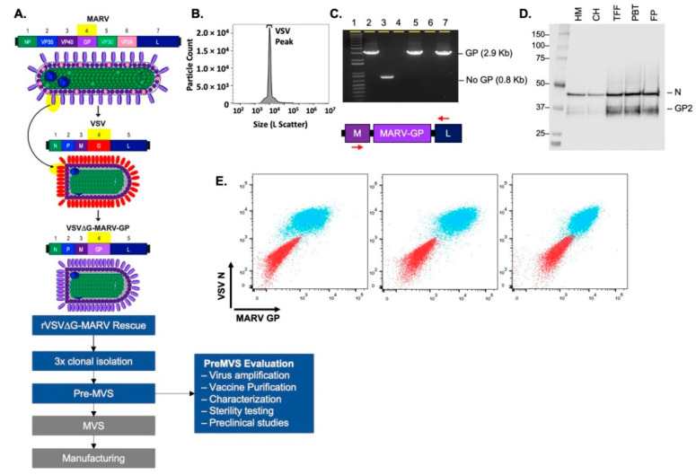
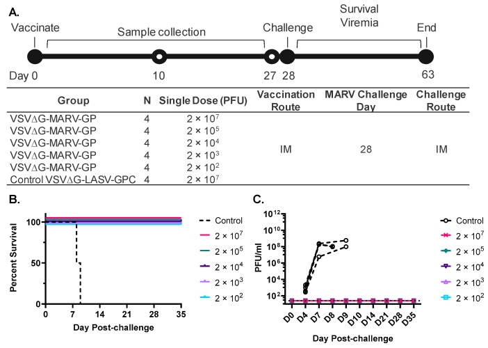
Vaccines are needed to disrupt or prevent continued outbreaks of filoviruses in humans across Western and Central Africa, including outbreaks of Marburg virus (MARV). As part of a filovirus vaccine product development plan, it is important to investigate dose response early in preclinical development to identify the dose range that may be optimal for safety, immunogenicity, and efficacy, and perhaps demonstrate that using lower doses is feasible, which will improve product access. To determine the efficacious dose range for a manufacturing-ready live recombinant vesicular stomatitis virus vaccine vector (rVSV∆G-MARV-GP) encoding the MARV glycoprotein (GP), a dose-range study was conducted in cynomolgus macaques. Results showed that a single intramuscular injection with as little as 200 plaque-forming units (PFUs) was 100% efficacious against lethality and prevented development of viremia and clinical pathologies associated with MARV Angola infection. Across the vaccine doses tested, there was nearly a 2000-fold range of anti-MARV glycoprotein (GP) serum IgG titers with seroconversion detectable even at the lowest doses. Virus-neutralizing serum antibodies also were detected in animals vaccinated with the higher vaccine doses indicating that vaccination induced functional antibodies, but that the assay was a less sensitive indicator of seroconversion. Collectively, the data indicates that a relatively wide range of anti-GP serum IgG titers are observed in animals that are protected from disease implying that seroconversion is positively associated with efficacy, but that more extensive immunologic analyses on samples collected from our study as well as future preclinical studies will be valuable in identifying additional immune responses correlated with protection that can serve as markers to monitor in human trials needed to generate data that can support vaccine licensure in the future.

Keywords: Marburg virus vaccine; emerging infectious disease; filoviruses; vaccine vector; vesicular stomatitis virus.


Conflict of interest statement

C.L.C., C.L.P., M.Y., E.S., J.W.C. and M.B.F. have filed a provisional patent application on the Marburg virus vaccine candidate. The authors declare no other conflict of interest.

Figures

Figure A1
Figure A1
MARV RNA detected in blood after virus challenge. RNA was extracted from whole blood and was quantified by RT-qPCR. Dashed line represents LOD for assay of 1000 genome copies/mL.
Figure 1
Figure 1
Generation and characterization of the rVSV∆G-MARV-GP vaccine for use in humans. (A) The schematic summarizes the rVSV∆G-MARV-GP chimeric virus design and critical work stages leading to cGMP MVS production and vaccine manufacturing. Following the regeneration of a new recombinant rVSV∆G-MARV-GP virus, 3 rounds of virus plaque isolation were performed prior to the generation and evaluation of a pre-MVS. (B) Nanoflow cytometry using an Apogee instrument was performed [51] with rVSV∆G-MARV-GP virions purified using tangential flow filtration (TFF). The graph shows particle counts (Y axis) and large-angle light scatter (X-axis). Total particles for the vaccine candidate were 6.7 × 10¹⁰ particles/mL, VSV virus peak (indicated) corresponded to 73.2% of the total particles. (C) GP gene integrity was evaluated by RT-PCR using primers specific for the VSV M and L genes that flank the GP insert. Analysis of PCR products by agarose gel electrophoresis showed that the expected 2.9 Kb product was amplified and that no larger or small products were detectable. Samples analyzed on the gel include: (1) 1 Kb ladder; (2) positive control VSV-MARV genomic plasmid template; (3) a negative control VSV genomic plasmid DNA (pVSV-G5) in which the G gene was moved to the 5′ terminus of the genome and no transcription unit is present between M and L [52]; (4) A negative control containing no template nucleic acid in RT-PCR reaction; (5) Medium harvest containing rVSV∆G-MARV-GP released by infected cells; (6) Empty well; (7) Purified rVSV∆G-MARV-GP. (D) Western blot analysis conducted using samples from various stages of virus purification. Blot probed with anti-GP rabbit polyclonal antisera that bound to MARV GP2 and rabbit poly-clonal anti-VSV-N. Analyzed samples included: Harvested medium (HM); clarified harvest (CH); product concentrated by TFF (TFF); product post-benzonase treatment (PBT); and final product (FP). (E) Flow cytometry was performed using Vero cells infected with purified rVSVΔG-MARV-GP vaccine material. Overlay dot plots displaying co-expression of MARV GP (x-axis) and VSV-N (y-axis) on uninfected VERO cells as controls (red population) and VERO cells 40 h post infection (MOI 0.001) with rVSV∆G-MARV-GP (blue population). GP was detected with three different anti-GP Abs (Pan-Filovirus anti-GP mAb (left panel), murine mAb 5C1 (middle panel), or rabbit anti-GP pAb (right panel)), and intracellular VSV-N was detected with murine mAb 10G4.
Figure 2
Figure 2
Design of rVSV∆G-MARV-GP preclinical dose-range efficacy study conducted in cynomolgus macaques. (A) Timing of activities during the course of the study are shown at the top and a table summarizing study design is shown below. The control rVSV∆G-based Lassa virus vaccine material (rVSV∆G-LASV-GPC; [45,53]) was produced from a new recombinant strain (IAVI unpublished). (B) Animal survival after MARV Angola challenge. (C) MARV viremia quantified by plaque assay at the indicated timepoints post viral challenge. Limit of detection (LOD) = 25 PFU/mL.
Figure 3
Figure 3
Characterization of the antibody response to vaccination. (A) ELISA was performed using plates coated with a soluble form of MARV GP Angola. Day 27 post-vaccination endpoint titers plotted as mean and standard deviation with animal numbers corresponding to high responders are shown in the graph. Vaccine dose in PFUs is included at the right side of the graph. The dashed line represents the lower detection limit of assay of 100. Statistical analysis was performed comparing vaccine cohort responses to vector control vaccinated animals using an ANOVA multiple comparison (n = 4, * p ≤ 0.05, *** p ≤ 0.001). (B) Serum dilution of pre-immune (Pre) and Day 27 (D27) post-vaccination at which rVSV∆G-MARV-GP (Musoke) plaque numbers were reduced by 50% (Neutralization titer 50 or NT 50) is plotted. The dashed line represents the lower detection limit of assay, 20. N = 4, mean and standard deviation.

References

    1. Sun J., Uwishema O., Kassem H., Abbass M., Uweis L., Rai A., El Saleh R., Adanur I., Onyeaka H. Ebola virus outbreak returns to the Democratic Republic of Congo: An urgent rising concern. Ann. Med. Surg. 2022;79:103958. doi: 10.1016/j.amsu.2022.103958. - DOI - PMC - PubMed
    1. Keita A.K., Koundouno F.R., Faye M., Dux A., Hinzmann J., Diallo H., Ayouba A., Le Marcis F., Soropogui B., Ifono K., et al. Resurgence of Ebola virus in 2021 in Guinea suggests a new paradigm for outbreaks. Nature. 2021;597:539–543. doi: 10.1038/s41586-021-03901-9. - DOI - PubMed
    1. WHO Ebola Virus Disease—Democratic Republic of the Congo. [(accessed on 8 July 2022)]. Available online: https://www.who.int/emergencies/disease-outbreak-news/item/2022-DON377.
    1. Munster V.J., Bausch D.G., de Wit E., Fischer R., Kobinger G., Munoz-Fontela C., Olson S.H., Seifert S.N., Sprecher A., Ntoumi F., et al. Outbreaks in a Rapidly Changing Central Africa—Lessons from Ebola. N. Engl. J. Med. 2018;379:1198–1201. doi: 10.1056/NEJMp1807691. - DOI - PubMed
    1. Pigott D.M., Golding N., Mylne A., Huang Z., Weiss D.J., Brady O.J., Kraemer M.U., Hay S.I. Mapping the zoonotic niche of Marburg virus disease in Africa. Trans. R. Soc. Trop. Med. Hyg. 2015;109:366–378. doi: 10.1093/trstmh/trv024. - DOI - PMC - PubMed