. 2022 Sep 23;10(10):1603.
doi: 10.3390/vaccines10101603.

The Deletion of US3 Gene of Pseudorabies Virus (PRV) ΔgE/TK Strain Induces Increased Immunogenicity in Mice

Affiliations

The Deletion of US3 Gene of Pseudorabies Virus (PRV) ΔgE/TK Strain Induces Increased Immunogenicity in Mice

Meng-Meng Deng et al. Vaccines (Basel). .

Abstract

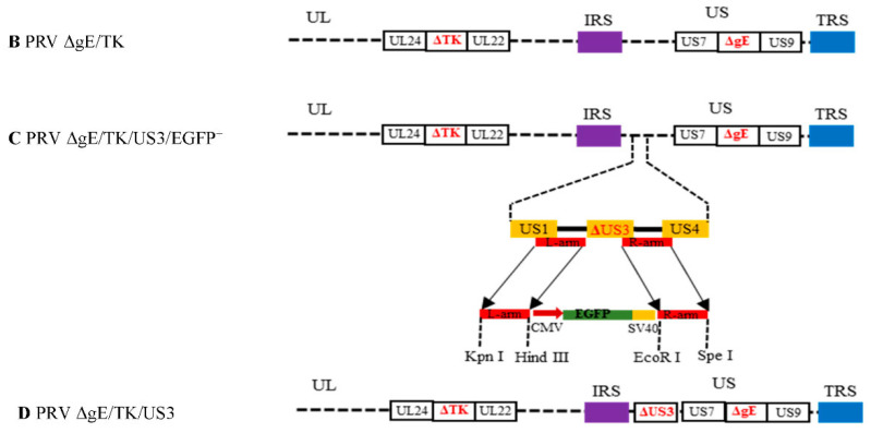
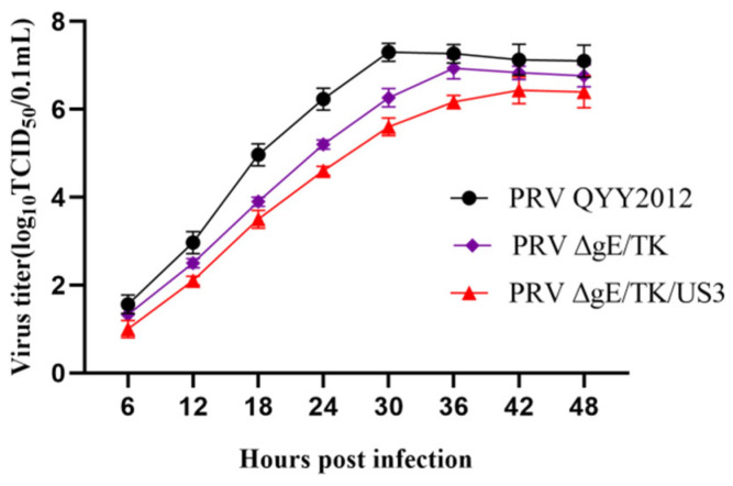
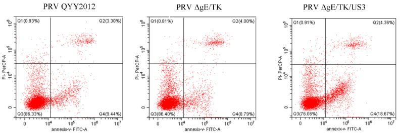
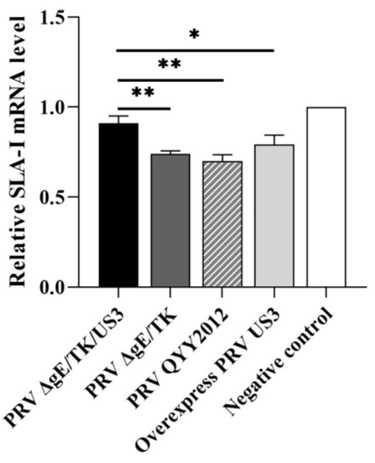
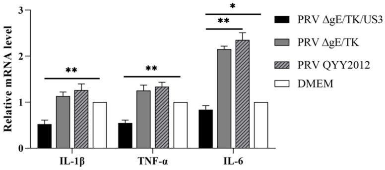
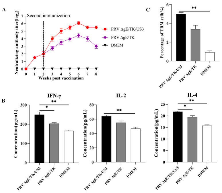
Re-emerging pseudorabies (PR) caused by pseudorabies virus (PRV) variant has been prevailing among immunized herds in China since 2011, indicating that commercially available PR vaccine strains couldn't provide complete protection against novel, epidemic PRV variant. Before this study, a gE/TK-gene-deleted virus (PRV ΔgE/TK) was constructed from PRV QYY2012 variant through homologous recombination and Cre/LoxP system. Here, PRV ΔgE/TK/US3 strain was generated by deleting US3 gene based on PRV ΔgE/TK strain using the same method. The growth characteristics of PRV ΔgE/TK/US3 were analogous to that of PRV ΔgE/TK. Moreover, the deletion of US3 gene could promote apoptosis, upregulate the level of swine leukocyte antigen class I molecule (SLA-I) in vitro, and relieve inflammatory response in inoculated BALB/c mice. Subsequently, the safety and immunogenicity of PRV ΔgE/TK/US3 was evaluated as a vaccine candidate in mice. The results revealed that PRV ΔgE/TK/US3 was safe for mice, and mice vaccinated with PRV ΔgE/TK/US3 could induce a higher level of PRV-specific neutralizing antibodies and cytokines, including IFN-γ, IL-2 and IL-4, also higher level of CD8+ CD69+ Tissue-Resident Memory T cells (TRM). The results show that the deletion of US3 gene of PRV ΔgE/TK strain could induce increased immunogenicity, indicating that the PRV ΔgE/TK/US3 strain is a promising vaccine candidate for preventing and controlling of the epidemic PR in China.

Keywords: PRV ΔgE/TK/US3; US3 gene; immunogenicity; pseudorabies virus.


Conflict of interest statement

The authors declare no conflict of interest.

Figures

Figure 1
Figure 1
Schematic diagrams of PRV ΔgE/TK/US3. (A) pSK-US3-LR-EGFP recombinant plasmid. (B)The schematic representation of PRV ΔgE/TK genome in which partial coding regions of gE and TK genes are deleted. (C) The schematic representation of PRV ΔgE/TK/US3/EGFP+ genome in which partial coding regions of gE, TK and US3 genes are deleted and an EGFP expression cassette is inserted into the US3 deleted locus. (D) The schematic representation of PRV ΔgE/TK/US3 genome.
Figure 1
Figure 1
Schematic diagrams of PRV ΔgE/TK/US3. (A) pSK-US3-LR-EGFP recombinant plasmid. (B)The schematic representation of PRV ΔgE/TK genome in which partial coding regions of gE and TK genes are deleted. (C) The schematic representation of PRV ΔgE/TK/US3/EGFP+ genome in which partial coding regions of gE, TK and US3 genes are deleted and an EGFP expression cassette is inserted into the US3 deleted locus. (D) The schematic representation of PRV ΔgE/TK/US3 genome.
Figure 2
Figure 2
Purification and identification of PRV strains. (A) Plaque purification of PRV ΔgE/TK/US3/EGFP+ strain for fluorescence visual field. (B) Plaque purification of PRV ΔgE/TK/US3/EGFP+ strain for bright visual field, red circles represent the plaque of PRV ΔgE/TK/US3/EGFP+. (C) Plaque purification of PRV ΔgE/TK/US3 strain for fluorescence visual field. (D) Plaque purification of PRV ΔgE/TK/US3 strain for bright visual field, red circles represent the plaque of PRV ΔgE/TK/US3. (E) PCR analysis of different PRV strains with primers US3-F/R. Lane 1, PRV QYY2012; lane 2, PRV ΔgE/TK/US3/EGFP+; lane 3, PRV ΔgE/TK/US3; lane 4, negative control; M, DL2000 DNA Marker.
Figure 3
Figure 3
The multiple step growth curves of different PRV strains. Vero cells were inoculated with PRV QYY2012, PRV ΔgE/TK, or PRV ΔgE/TK/US3 strains. The cell culture supernatants were collected at different time points and used to calculate the TCID50 of each virus.
Figure 4
Figure 4
The levels of apoptosis of Vero cells infected with different PRV strains. Vero cells were inoculated with PRV QYY2012, PRV ΔgE/TK, or PRV ΔgE/TK/US3 strains. At 24 h after infection, cells were stained with Annexin V-FITC/PI and analyzed by flow cytometry.
Figure 5
Figure 5
The expression level of SLA-I in ST cells infected with different PRV strains.ST cells were inoculated with PRV QYY2012, PRV ΔgE/TK or PRV ΔgE/TK/US3 strains, and pUS3 plasmid was overexpressed in ST cells. At 6 h after infection or transfection, RNA in infected ST cells was extracted and detected by RT-qPCR using primers SLA-I qF/R. Data was presented as mean ± SD.* p < 0.05, ** p < 0.01.
Figure 6
Figure 6
The expression level of pro-inflammatory cytokines in mice brain tissues inoculated with different PRV strains. At 48 h after inoculation, RNA in mice brain tissues was extracted and detected by RT-qPCR using primers of IL-1β, TNF-α, and IL-6. Data was presented as mean ± SD.* p < 0.05, ** p < 0.01.
Figure 7
Figure 7
The immunogenicity of PRV ΔgE/TK/US3 in mice (A) Neutralizing antibody titer was examined in the serum of vaccinated mice. (B) The expression levels of cytokines IFN-γ, IL-2, and IL-4 in the peripheral serum of vaccinated mice were detected by ELISA. (C) The number of CD8+ CD69+ TRM cells in trigeminal ganglion of vaccinated mice was detected by flow cytometry. Data was presented as mean ± SD.* p < 0.05, ** p < 0.01.

References

    1. Freuling C.M., Müller T.F., Mettenleiter T.C. Vaccines against pseudorabies virus (PrV) Vet. Microbiol. 2017;206:3–9. doi: 10.1016/j.vetmic.2016.11.019. - DOI - PubMed
    1. Zhao H., Wang S., Liu C., Han J., Tang J., Zhou L., Ge X., Guo X., Yang H. The pUL56 of pseudorabies virus variant induces downregulation of swine leukocyte antigen class I molecules through the lysosome pathway. Virus Res. 2018;251:56–67. doi: 10.1016/j.virusres.2018.04.007. - DOI - PubMed
    1. Müller T., Hahn E.C., Tottewitz F., Kramer M., Klupp B.G., Mettenleiter T.C., Freuling C. Pseudorabies virus in wild swine: A global perspective. Arch. Virol. 2011;156:1691–1705. doi: 10.1007/s00705-011-1080-2. - DOI - PubMed
    1. Laval K., Enquist L.W. The Neuropathic Itch Caused by Pseudorabies Virus. Pathogens. 2020;9:254. doi: 10.3390/pathogens9040254. - DOI - PMC - PubMed
    1. Ao J.Q., Wang J.W., Chen X.H., Wang X.Z., Long Q.X. Expression of pseudorabies virus gE epitopes in Pichia pastoris and its utilization in an indirect PRV gE-ELISA. J. Virol. Methods. 2003;114:145–150. doi: 10.1016/j.jviromet.2003.09.012. - DOI - PubMed