. 2022 Oct 10;10(10):1692.
doi: 10.3390/vaccines10101692.

Recurrent and De Novo Toxoplasmosis Retinochoroiditis following Coronavirus Disease 2019 Infection or Vaccination

Affiliations

Recurrent and De Novo Toxoplasmosis Retinochoroiditis following Coronavirus Disease 2019 Infection or Vaccination

Mélanie Hébert et al. Vaccines (Basel). .

Abstract

This study reports three cases of toxoplasmosis retinochoroiditis following coronavirus disease 2019 (COVID-19) infection or vaccination from the national Canadian COVID-19 Eye Registry between December 2020 and September 2021. A 56-year-old male presented 15 days after a positive COVID-19 test with toxoplasmosis retinochoroiditis. He later relapsed 8 days following a first Pfizer-BioNTech vaccine dose. Two patients presented with toxoplasmosis retinochoroiditis following COVID-19 vaccination: A 58-year-old female presenting 4 days following a first Pfizer-BioNTech vaccine dose with anterior uveitis and a posterior pole lesion discovered 3 months later and a 39-year-old female presenting 17 days after a first Moderna vaccine dose. Resolution was achieved with oral clindamycin, oral trimethoprim/sulfamethoxazole, and topical prednisolone acetate 1%. Patients were offered prophylactic trimethoprim/sulfamethoxazole for subsequent doses without relapse. Following COVID-19 infection or vaccination, patients may be at risk for toxoplasmosis retinochoroiditis. Prophylactic antibiotics for future doses may be offered to patients with known ocular toxoplasmosis to prevent recurrence.

Keywords: COVID-19 vaccination; SARS-CoV-2; antibiotic prophylaxis; coronavirus disease 2019; inflammation; mRNA vaccine; ophthalmic adverse events; toxoplasmosis retinochoroiditis; vaccination.


Conflict of interest statement

The authors declare no conflict of interest.

Figures
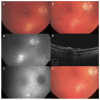
Figure 1
Figure 1
Case 1 with active toxoplasmosis retinochoroiditis following a diagnosis of coronavirus disease 2019 (COVID-19) and recurrence after COVID-19 vaccination. (A) Color fundus photography showing a single, white, deep chorioretinal lesion along the inferior arcade with vitritis at initial presentation. (B) Initial red-free monochromatic fundus photograph showing the same lesion which becomes more apparent. (C) Initial fundus autofluorescence showing a hyperautofluorescent lesion with distinct borders corresponding to the retinochoroiditis. (D) Four months after presentation, the lesion is now cicatricial without vitritis with (E) a horizontal optical coherence tomography scan through the lesion. (F) The patient had a recurrence 1 month later, 8 days after his first COVID-19 vaccine dose.
Figure 2
Figure 2
Case 2 with active toxoplasmosis retinochoroiditis 3 months after a first dose of COVID-19 vaccination. (A) Fundus photography showing the initial elevated, yellow whitish lesion with surrounding pigmentary changes. (B) Six weeks later, the main lesion became cicatricial.
Figure 3
Figure 3
Case 3 with a previous, cicatricial toxoplasmosis scar with recurrence of active toxoplasmosis retinochoroiditis 17 days after COVID-19 vaccination. (A) Fundus photography showcasing the old toxoplasmosis scar with an area of active retinochoroiditis adjacent to it. There is diffuse sheathing consistent with vasculitis. (B) Three weeks later, the new area of retinochoroiditis has decreased in size and activity as did the vasculitis. (C) At seven weeks, there was still an area of activity within the chorioretinal scar, thus the treatment was further extended. (D) At final follow-up 4 months later, the area had scarred, and no inflammation was observed.

References

    1. Smith J.R., Ashander L.M., Arruda S.L., Cordeiro C.A., Lie S., Rochet E., Belfort R., Jr., Furtado J.M. Pathogenesis of ocular toxoplasmosis. Prog. Retin. Eye Res. 2021;81:100882. doi: 10.1016/j.preteyeres.2020.100882. - DOI - PubMed
    1. Vishnevskia-Dai V., Achiron A., Buhbut O., Berar O.V., Musika A.A., Elyashiv S.M., Hecht I. Chorio-retinal toxoplasmosis: Treatment outcomes, lesion evolution and long-term follow-up in a single tertiary center. Int. Ophthalmol. 2020;40:811–821. doi: 10.1007/s10792-019-01242-1. - DOI - PubMed
    1. Olson D.J., Parhiz A.T., Wirthlin R.S. Reactivation of Latent Toxoplasmosis Following Dexamethasone Implant Injection. Ophthalmic Surg. Lasers Imaging Retin. 2016;47:1050–1052. doi: 10.3928/23258160-20161031-10. - DOI - PubMed
    1. Puga M., Carpio D., Sampil M., Zamora M.J., Fernandez-Salgado E. Ocular Toxoplasmosis Reactivation in a Patient With Inflammatory Bowel Disease Under Treatment With Azathioprine. J. Clin. Gastroenterol. 2016;50:610. doi: 10.1097/MCG.0000000000000521. - DOI - PubMed
    1. Zhou R., To K.K., Wong Y.C., Liu L., Zhou B., Li X., Huang H., Mo Y., Luk T.Y., Lau T.T., et al. Acute SARS-CoV-2 Infection Impairs Dendritic Cell and T Cell Responses. Immunity. 2020;53:864–877.e5. doi: 10.1016/j.immuni.2020.07.026. - DOI - PMC - PubMed

LinkOut - more resources