. 2022 Sep 23;14(10):2109.
doi: 10.3390/v14102109.

Chimeric Virus-like Particles Co-Displaying Hemagglutinin Stem and the C-Terminal Fragment of DnaK Confer Heterologous Influenza Protection in Mice

Affiliations

Chimeric Virus-like Particles Co-Displaying Hemagglutinin Stem and the C-Terminal Fragment of DnaK Confer Heterologous Influenza Protection in Mice

Cui-Cui Liu et al. Viruses. .

Abstract

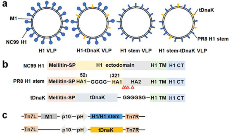
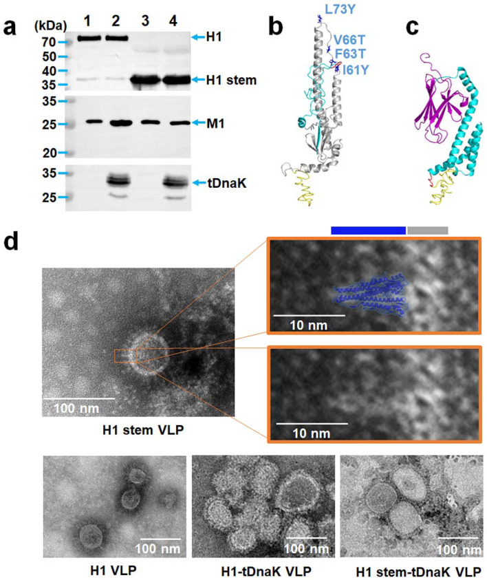
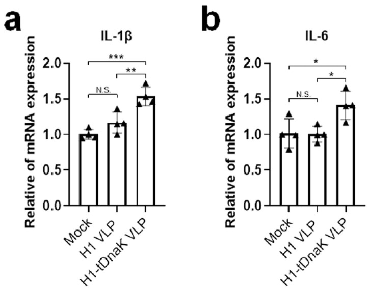
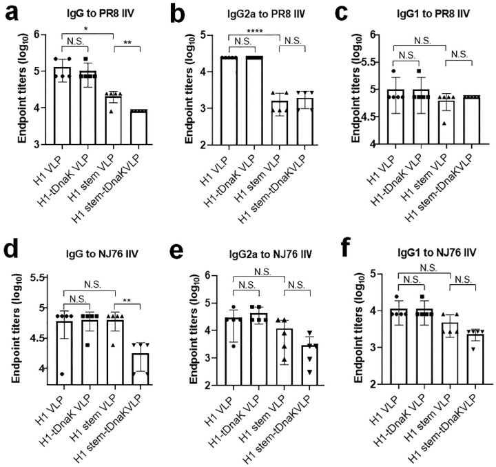
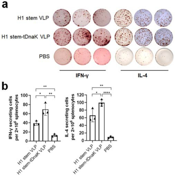
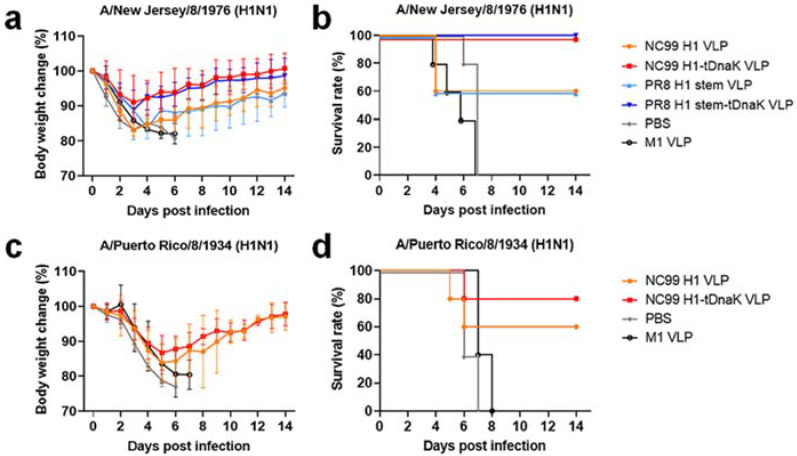
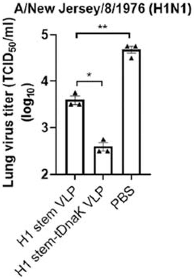
Influenza virus hemagglutinin (HA) stem is currently regarded as an extremely promising immunogen for designing universal influenza vaccines. The appropriate antigen-presenting vaccine vector would be conducive to increasing the immunogenicity of the HA stem antigen. In this study, we generated chimeric virus-like particles (cVLPs) co-displaying the truncated C-terminal of DnaK from Escherichia coli and H1 stem or full-length H1 antigen using the baculovirus expression system. Transmission electronic micrography revealed the expression and presentation of H1 stem antigens on the surface of VLPs. Vaccinations of mice with the H1 stem cVLPs induced H1-specific immune responses and provided heterologous immune protection in vivo, which was more effective than vaccinations with VLPs displaying H1 stem alone in protecting mice against weight loss as well as increasing survival rates after lethal influenza viral challenge. The results indicate that the incorporation of the truncated C-terminal of DnaK as an adjuvant protein into the cVLPs significantly enhances the H1-specific immunity and immune protection. We have explicitly identified the VLP platform as an effective way of expressing HA stem antigen and revealed that chimeric VLP is an vaccine vector for developing HA stem-based universal influenza vaccines.

Keywords: DnaK; hemagglutinin stem; influenza virus; universal vaccine; virus-like particle.


Conflict of interest statement

The authors declare no conflict of interest.

Figures

Figure 1
Figure 1
The construction of the cVLPs. (a) Schematic diagrams of the chimeric H1 VLP, H1-tDnaK VLP, H1 stem VLP, and H1 stem-tDnaK VLP. (b) Schematic diagrams of the designs of the full-length NC99 H1, PR8 H1 stem, and membrane-anchored tDnaK. Red arrows indicate site-mutations, I61Y, F63T, V66T, and L73Y in PR8 HA2. (c) Schematic diagrams of the designs of recombinant pFastBac Dual plasmids encoding M1 and H1/H1 stem, or membrane-anchored truncated DnaK alone. These exogenous genes were cloned into two independent open reading frames under the controls of the baculovirus polyhedrin promoter (pH) and the p10 promoter, respectively. All the cVLPs were purified by using density-gradient ultra-centrifugation. Western blotting analysis using recombinant H1 (NC99 H1N1)-specific mouse sera, the M1-specific monoclonal antibody C111, and DnaK-specific polyclonal antibodies clearly showed the presence of H1 and M1 in the H1 VLPs, H1, M1, and tDnaK in the H1-tDnaK VLPs, the H1 stem and M1 in the H1 stem VLPs, the H1 stem, M1 and tDnaK in the H1 stem-tDnaK VLPs (Figure 2a). To evaluate the size and conformation of H1 stem and tDnaK recombinant proteins, the structures of these two proteins were successfully predicted using the I-TASSER server (Figure 2b,c). Transmission electron micrography clearly showed the cVLP of approximately 80 nm in size and the good presentation of H1 stem antigens on the VLP membrane (Figure 2d). A cartoon image of the intercepted H1 stem (PDB: 1RU7) generated by using PyMOL was overlain on a spike in the TEM image with the scale bar of the same size and was found to be quite similar in size and shape to the spike on the VLP surface. According to the image superimposition analysis, we speculate that the intact fusion peptide and H1 transmembrane domain immobilize the H1 stem pre-fusion trimeric state.
Figure 2
Figure 2
The characterization of the cVLPs. (a) Western blot analysis of the expressions of the full-length H1, H1 stem, M1, and tDnaK from the cVLPs, including (1) H1 VLPs, (2) H1-tDnaK VLPs, (3) H1 stem VLPs, and (4) H1 stem-tDnaK VLPs. The predicted structures of (b) the PR8 H1 stem and (c) tDnaK from E. coli. (d) Transmission electron microscope analysis of H1 stem VLP, H1 stem-tDnaK VLP, H1 VLP, or H1-tDnaK VLP. White bars represent 100 nm in the chimeric VLP images and 10 nm in the images of partial enlargement. The blue and grey bars above the right image indicate the lengths of the H1 stem domain and lipid layer. The cartoon of the H1 stem that was intercepted from the PR8 H1 structure (PDB: 1RU7) was generated using PyMOL software. The shape and size were compared by image overlay with TEM image and cartoon image. The height of the H1 stem in the cartoon measured by using PyMOL is approximately 10 nm.
Figure 3
Figure 3
Analysis of the biological activity of membrane-anchored C-terminal domain of DnaK in vitro. Human lung carcinoma cell line A549 cell cultures were mock-treated with PBS or treated with NC99 H1 VLPs and NC99 H1-tDnaK VLPs, respectively. Cell cultures were collected 24 h after treatment for transcription analysis, and then total RNA from cell samples was extracted and reverse-transcribed into cDNA immediately. The gene transcription levels of (a) IL-1β and (b) IL-6 were quantified by qPCR. The results were presented as means with standard deviation (n = 4 per group). Each triangle symbol represents the relative value of cytokine mRNA expression level of each VLP-treated or mock-treated cell culture sample. Statistical significance was analyzed by t-test; *, **, and *** represent p < 0.05, p < 0.01, and p < 0.001, respectively. N.S. indicates no significance.
Figure 4
Figure 4
Immunization of mice with the chimeric VLPs’ results in specific serum IgG responses. The error bars represent standard deviations. (ac) PR8-specific and (df) NJ76-specific serum antibody titers of (a,d) total IgG, (b,e) IgG2a, and (c,f) IgG1 were determined by using ELISA. Antibody endpoint titer is defined as the reciprocal of the highest dilution of a serum that gives a value of OD (450 nm) two-fold above the value of the pre-immune serum negative control. The results are presented as means with standard deviation (n = 5 per group). Each symbol represents the serum antibody endpoint titer of each mouse. Statistical significance was analyzed by t-test, *, **, and **** represent p < 0.05, p < 0.01, and p < 0.001, respectively. N.S. indicates no significance.
Figure 5
Figure 5
ELISPOT analysis of cellular immune responses. Mouse splenocyte samples (n = 3 per group) were collected on day 4 post sublethal-dose infections with 0.2 × LD50 of the NJ76 H1N1 virus. (a) Inactivated NJ76 H1N1 influenza virus re-stimulated IFN-γ- and IL-4-secreting cells were determined using ELISPOT assays. (b) The spot numbers of inactivated H1N1 virus-restimulated groups were quantified and compared in bar charts. The results were presented as means ± standard deviation. Each symbol represents the average spot number of the parallel samples from each mouse. The statistical significance was analyzed by t-test. *, **, and **** represent p < 0.05, p < 0.01, and p < 0.0001, respectively. N.S. indicates no significance.
Figure 6
Figure 6
VLP protective efficacy in BALB/c mice. Body weight changes (a,c) and survival rates (b,d) of the immunized mice (n = 5 per group) upon challenges with 3 × LD50 lethal dose infections with NJ76 (a,b) or PR8 H1N1 (c,d).
Figure 7
Figure 7
The determination of lung viral load after the sublethal infection. The purpose of the infection experiment is to compare the in vivo protectiveness among vaccination groups; the sublethal dose of mouse-adapted NJ76 was employed for infection for the consideration of lab-animal welfare. Mouse lung samples (n = 3 per group) were collected on day 4 post sublethal-dose infections with 1 × LD50 NJ76 H1N1 virus. Viral titers are expressed as TCID50/mL. The bars present the mean value ± standard deviation. Each symbol represents the average virus titer of the parallel samples from each mouse. Statistical significance was analyzed by t-test. * represents p < 0.05, and ** represents p < 0.01.

Similar articles

Cited by

References

    1. Becker T., Elbahesh H., Reperant L.A., Rimmelzwaan G.F., Osterhaus A.D.M.E. Influenza Vaccines: Successes and Continuing Challenges. J. Infect. Dis. 2021;224:S405–S419. doi: 10.1093/infdis/jiab269. - DOI - PMC - PubMed
    1. Krammer F., Smith G.J.D., Fouchier R.A.M., Peiris M., Kedzierska K., Doherty P.C., Palese P., Shaw M.L., Treanor J., Webster R.G., et al. Influenza. Nat. Rev. Dis. Primers. 2018;4:3. doi: 10.1038/s41572-018-0002-y. - DOI - PMC - PubMed
    1. Wei C.J., Crank M.C., Shiver J., Graham B.S., Mascola J.R., Nabel G.J. Next-generation influenza vaccines: Opportunities and challenges. Nat. Rev. Drug Discov. 2020;19:239–252. doi: 10.1038/s41573-019-0056-x. - DOI - PMC - PubMed
    1. Kosik I., Yewdell J.W. Influenza Hemagglutinin and Neuraminidase: Yin–Yang Proteins Coevolving to Thwart Immunity. Viruses. 2019;11:346. doi: 10.3390/v11040346. - DOI - PMC - PubMed
    1. Kosik I., Ince W.L., Gentles L.E., Oler A.J., Kosikova M., Angel M., Magadan J.G., Xie H., Brooke C.B., Yewdell J.W. Influenza A virus hemagglutinin glycosylation compensates for antibody escape fitness costs. PLoS Pathog. 2018;14:e1006796. - PMC - PubMed

Publication types

Substances