. 2022 Sep 30;14(10):2174.
doi: 10.3390/v14102174.

Characterization and Pathogenicity of Two Novel PRRSVs Recombined by NADC30-like and NADC34-like Strains in China

Affiliations

Characterization and Pathogenicity of Two Novel PRRSVs Recombined by NADC30-like and NADC34-like Strains in China

Yu Wu et al. Viruses. .

Abstract

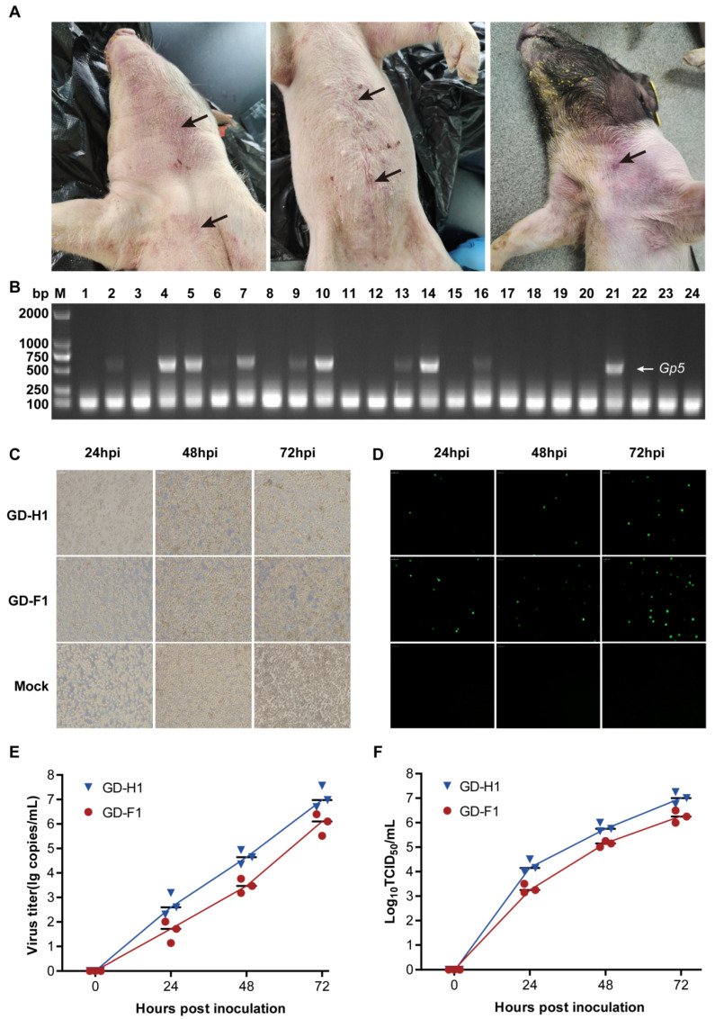
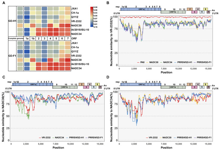
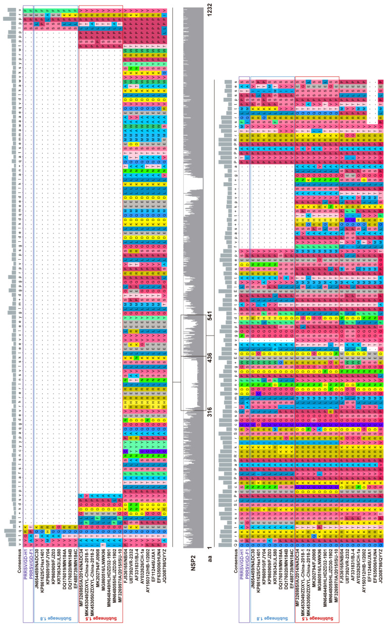
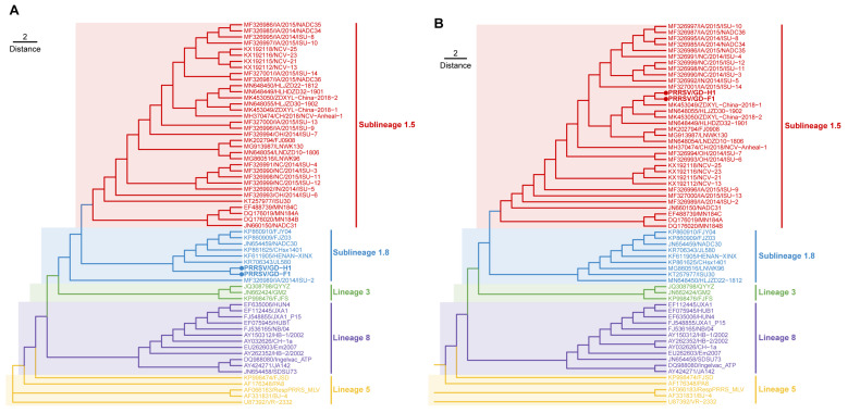
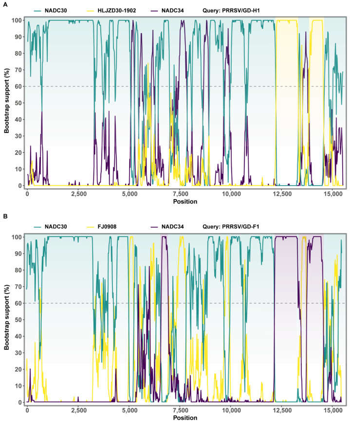
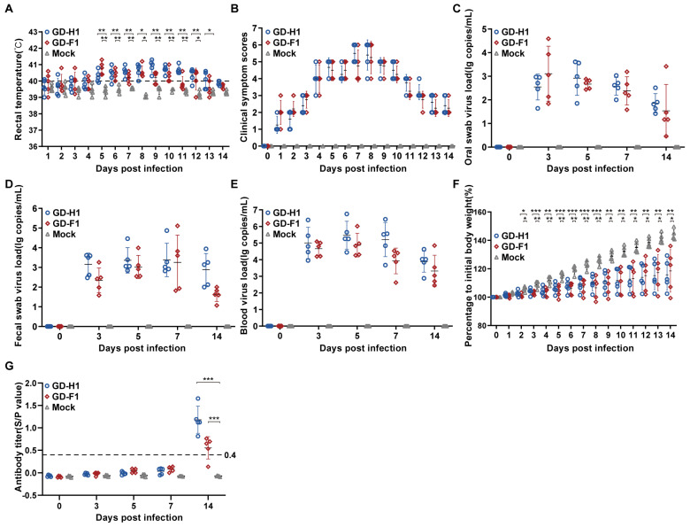
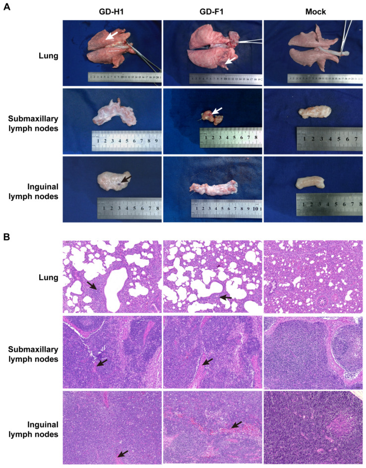
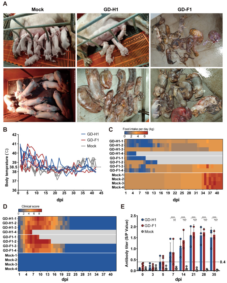
Porcine reproductive and respiratory syndrome viruses (PRRSVs) pose a serious threat to the swine industry in China, which has caused great difficulties for porcine reproductive and respiratory syndrome (PRRS) immune prevention and control, due to its easily mutable and recombinant nature. In this study, two novel PRRSV strains, which were named GD-H1 and GD-F1, were isolated and fully sequenced from pig farms in Guangdong province, China. The phylogenetic analysis and recombination analysis revealed that the GD-H1 and GD-F1 were generated by the recombination of NADC30-like and NADC34-like strains which were different from the previously prevalent strain. Further pathogenic studies on piglets and sows found that the recombinant strains could cause piglets high fever, loss of appetite and lung lesions, but no piglets died. However, the recombinant strains could cause acute death and abortion in pregnant sow infection models together with average survival rates of 62.5% and 37.5% abortion rates, respectively. These findings indicated that the recombinant strains were extremely pathogenic to sows. Therefore, we report two clinical novel recombinant strains of PRRSV that are different from the traditional epidemic strains in China, which may provide early warning and support for PRRS immune prevention and control.

Keywords: PRRSV; pathogenicity; phylogenetic analyses; recombination analyses.


Conflict of interest statement

The authors declare no potential conflict of interest with respect to the research, authorship and publication of this article.

Figures

Figure 1
Figure 1
Strain isolation and identification. (A) Clinical symptoms of PRRSV-infected pigs; black arrows mark the lesion sections. (B) Clinical samples were examined by gel electrophoresis. (C) Cytopathic conditions at 24, 36, and 72 h post-infection (hpi). (D) IFAs showing the reactivity of a monoclonal antibody against PRRSV N protein to GD-H1 and GD-F1 strains infected at 24, 36, and 72 hpi. Magnification, 400×. (E) Viral titer detection of GD-H1 and GD-F1 strains at 24, 36, and 72 hpi. (F) Growth kinetic of GD-H1 and GD-F1 strains.
Figure 2
Figure 2
GD-H1 and GD-F1 strains’ amino acid similarity to classical strains. (A) heatmap compared the similarities of isolated strains and other reference strains. (BD) Comparison of GD-H1 and GD-F1 strains’ similarity to VR-2332, NADC30, and NADC34 strains.
Figure 3
Figure 3
Alignment of the deduced amino acid sequence based on the NSP2 gene. The alignment of the GD-H1 and GD-F1 strains is marked with purple.
Figure 4
Figure 4
Phylogenetic and recombination analysis of GD-H1 and GD-F1 strains. (A) Phylogenetic trees constructed based on the ORF5 gene of GD-H1 and GD-F1 strains with 61 reference PRRSV strains. (B) Phylogenetic trees constructed based on the full-length genomes of the GD-H1 and GD-F1 strains with 61 reference PRRSV strains. The GD-H1 and GD-F1 strains are marked with a round dot. The NADC34-like and NADC30-like isolates are labeled in red and blue.
Figure 5
Figure 5
The crossover regions in the GD-H1 and GD-F1 genomes were further confirmed by SimPlot 3.5.1. (A) For GD-H1, NADC30 is the major parental virus while HLJZD30-1902 as the minor parental viruses. (B) For GD-F1, NADC30 is the major parental virus while NADC34 as the minor parental viruses. The crossover regions identified by SimPlot were consistent with the results from RDP4 analysis (Table S4). The y-axis shows the percentage of permutated trees employing a sliding window of 200 nucleotides (nt) and a step size of 20 nt.
Figure 6
Figure 6
Pathogenicity results in piglets. (A) Body temperature change of pigs in each group after challenge. (B) Clinical scores of pigs after challenge during the entire experiment. (CE) Viral load detection in blood, oral swabs and pharyngeal swabs. (F) Body weight gains of each group during the challenge study. (G) PRRSV-specific antibody level was detected in each group during the challenge study. Each bar represents the average for all pigs in each group ± standard deviation (SD). The significant difference is marked with the asterisk, *** p < 0.001, ** p < 0.01, and * p < 0.05.
Figure 7
Figure 7
Observation and detection by pathological necropsy. (A) Necropsy observation of lung, submandibular lymph nodes and inguinal lymph nodes. White arrows mark the site of tissue bleeding. (B) Histopathology tests of the lung, submandibular lymph nodes and inguinal lymph nodes. Black arrows mark the lung lesions site in HE straining sections, as well as lymph node bleeding lesions.
Figure 8
Figure 8
Pathogenicity results in pregnant sows. (A) Clinical photographs of the sow farrowing. (B) Body temperature change of pregnant sows in each group after challenge. (C) Food intake changes of pregnant sows in each group after challenge. (D) Clinical scores of pregnant sows after challenge during the entire experiment. (E) PRRSV-specific antibody level was detected in each group during the challenge study. Each bar represents the average for all pigs in each group ± standard deviation (SD). The significant difference is marked with the asterisk, *** p < 0.001 and ** p < 0.01.

Similar articles

Cited by

References

    1. Lole K.S., Bollinger R.C., Paranjape R.S., Gadkari D., Kulkarni S.S., Novak N.G., Ingersoll R., Sheppard H.W., Ray S.C. Full-length human immunodeficiency virus type 1 genomes from subtype C-infected seroconverters in India, with Evidence of Intersubtype Recombination. J. Virol. 1999;73:152–160. doi: 10.1128/JVI.73.1.152-160.1999. - DOI - PMC - PubMed
    1. Gao J.-C., Xiong J.-Y., Ye C., Chang X.-B., Guo J.-C., Jiang C.-G., Zhang G.-H., Tian Z.-J., Cai X.-H., Tong G.-Z., et al. Genotypic and geographical distribution of porcine reproductive and respiratory syndrome viruses in mainland China in 1996–2016. Vet. Microbiol. 2017;208:164–172. doi: 10.1016/j.vetmic.2017.08.003. - DOI - PubMed
    1. Firth A.E., Zevenhoven-Dobbe J.C., Wills N.M., Go Y.Y., Balasuriya U.B.R., Atkins J., Snijder E.J., Posthuma C.C. Discovery of a small arterivirus gene that overlaps the GP5 coding sequence and is important for virus production. J. Gen. Virol. 2011;92:1097–1106. doi: 10.1099/vir.0.029264-0. - DOI - PMC - PubMed
    1. Liu J., Wei C., Lin Z., Xia W., Ma Y., Dai A., Yang X. Full genome sequence analysis of a 1-7-4-like PRRSV strain in Fujian Province, China. PeerJ. 2019;7:e7859. doi: 10.7717/peerj.7859. - DOI - PMC - PubMed
    1. Thaa B., Sinhadri B.C., Tielesch C., Krause E., Veit M. Signal Peptide Cleavage from GP5 of PRRSV: A Minor Fraction of Molecules Retains the Decoy Epitope, a Presumed Molecular Cause for Viral Persistence. PLoS ONE. 2013;8:e65548. doi: 10.1371/journal.pone.0065548. - DOI - PMC - PubMed

Publication types

LinkOut - more resources