. 2022 Oct 19;14(10):2298.
doi: 10.3390/v14102298.

Investigation of the Association between the Energy Metabolism of the Insect Vector Laodelphax striatellus and Rice Stripe Virus (RSV)

Affiliations

Investigation of the Association between the Energy Metabolism of the Insect Vector Laodelphax striatellus and Rice Stripe Virus (RSV)

Lu Zhang et al. Viruses. .

Abstract

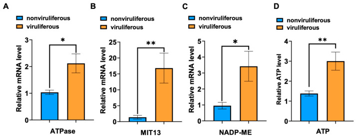
Viruses, as intracellular parasites, rely on the host organism to complete their life cycle. Although over 70% of plant viruses are transmitted by insect vectors, the role of vector energy metabolism on the infection process of insect-borne plant viruses is unclear. In this study, full-length cDNAs of three energy metabolism-related genes (LsATPase, LsMIT13 and LsNADP-ME) were obtained from the small brown planthopper (SBPH, Laodelphax striatellus), which transmits the Rice stripe virus (RSV). Expression levels of LsATPase, LsMIT13 and LsNADP-ME increased by 105%, 1120% and 259%, respectively, due to RSV infection. The repression of LsATPase, LsMIT13 or LsNADP-ME by RNAi had no effect on RSV nucleocapsid protein (NP) transcripts or protein levels. The repression of LsATPase caused a significant increase in LsMIT13 and LsNADP-ME transcript levels by 230% and 217%, respectively, and the repression of LsMIT13 caused a significant increase in LsNADP-ME mRNA levels. These results suggested that the silencing of LsATPase induced compensatory upregulation of LsMIT13 and LsNADP-ME, and silencing LsMIT13 induced compensatory upregulation of LsNADP-ME. Further study indicated that the co-silencing of LsATPase, LsMIT13 and LsNADP-ME in viruliferous SBPHs increased ATP production and RSV loads by 182% and 117%, respectively, as compared with nonviruliferous SBPHs. These findings indicate that SBPH energy metabolism is involved in RSV infection and provide insight into the association between plant viruses and energy metabolism in the insect vector.

Keywords: ATP synthase; Laodelphax striatellus; NAD-dependent malic enzyme; RSV; energy metabolism; mitochondrial import inner membrane translocases.


Conflict of interest statement

The authors declare no conflict of interest.

Figures

Figure 1
Figure 1
LsATPase, LsMIT13 and LsNADP-ME protein structure and amino acid alignment. Schematic representation and deduced amino acid alignments are shown for (A) ATPase, (B) MIT13 and (C) NADP-ME. The ATP-synthase domains, Tim10_DDP domain, malic domain and malic-M domain are indicated by blue, green, red and yellow boxes, respectively. Abbreviations indicate protein from the following insect species: Ls, Laodelphax striatellus; Nl, Nilaparvata lugens; Bt, Bombus terrestris; Dm, Drosophila melanogaster; and Bg, Blattella germanica. Alignments were constructed using Clustal W software. Blue shading indicates conserved ATPase residues; orange or grey shading indicates species-specific residues.
Figure 2
Figure 2
RSV infection increased ATP levels and the expression of LsATPase, LsMIT13 and LsNADP-ME in SBPH. Expression levels in viruliferous and nonviruliferous SBPH adults were obtained by RT-qPCR; panels show expression of (A) LsATPase, (B) LsMIT13 and (C) LsNADP-ME. Five insects comprised a single replicate, and each treatment contained three replicates. (D) ATP levels in viruliferous and nonviruliferous SBPH as determined by ATP bioluminescence assays. Significant differences were obtained using the Student’s t-test (* p < 0.05 and ** p < 0.01; means ± SEM).
Figure 3
Figure 3
Knockdown of LsATPase, LsMIT13 and LsNADP-ME expression and effects on RSV load in SBPH. Panels: (A) LsATPase expression in dsLsATPase-treated SBPH; (B) LsMIT13 expression in dsLsMIT13-treated SBPH; and (C) LsNADP-ME expression in dsLsNADP-ME-treated SBPH; dsGFP was used as a control. Each treatment contained three replicates, and significant differences were determined using the Student’s t-test (* p < 0.05 and ** p < 0.01; means ± SEM). (D) Western blot analysis of RSV NP in SBPH (n = 30) treated with dsLsATPase, dsLsMIT13, dsLsNADP-ME and dsGFP. Antisera for glyceraldehyde-3-phosphate dehydrogenase (GAPDH) was used as control. Immunofluorescence is shown for ovaries (panel E), salivary glands (F) and midguts (G) of viruliferous SBPH treated with dsLsATPase, dsLsMIT13, dsLsNADP-ME and dsGFP. Tissues were immunolabeled with anti-RSV NP (Alexa Fluor 488, green), stained with DAPI (blue) and examined by confocal microscopy. Each treatment was replicated five times. Bar = 50 µm.
Figure 4
Figure 4
Compensatory regulation of LsATPase, LsMIT13 and LsNADP-ME during RNAi knockdown experiments. Panels show expression levels of LsATPase, LsMIT13 and LsNADP-ME in (A) dsLsATPase-, (B) dsLsMIT13- and (C) dsLsNADP-ME-treated SBPHs. Each treatment contained three replicates. (D) ATP levels in dsLsATPase-, dsLsMIT13-, dsLsNADP-ME- and dsGFP-treated SBPHs as determined by an ATP bioluminescence assay. Significant differences were evaluated using the Student’s t-test (* p < 0.05 and ** p < 0.01; means ± SEM).
Figure 5
Figure 5
The effects of co-silencing LsATPase, LsMIT13 and LsNADP-ME on RSV loads and ATP content. (A) qRT-PCR analysis of LsATPase, LsMIT13, LsNADP-ME and RSV NP expression in SBPH co-silenced with dsLsATPase, dsLsMIT13 and dsLsNADP-ME and dsGFP. (B) Western blot analysis of RSV NP in co-silenced (dsLsATPase, dsLsMIT13 and dsLsNADP-ME) and dsGFP-treated SBPH. A total of 30 treated SBPH were used for protein extraction. Glyceraldehyde-3-phosphate dehydrogenase (GAPDH) was used as control. (C) ATP levels in co-silenced (dsLsATPase, dsLsMIT13 and dsLsNADP-ME) and dsGFP-treated viruliferous SBPH as determined with the ATP bioluminescence assay. (D) ATP levels in co-silenced and dsGFP-treated nonviruliferous SBPH as measured with the ATP bioluminescence assay. Significant differences were determined with the Student’s t-test (* p < 0.05).

References

    1. Yang Q., Catalano C.E. ATP serves as a nucleotide switch coupling the genome maturation and packaging motor complexes of a virus assembly machine. Nucleic Acids Res. 2020;48:5006–5015. doi: 10.1093/nar/gkaa205. - DOI - PMC - PubMed
    1. Zhang J., Jia L., Tsang C.M., Tsao S.W. Infectious Agents Associated Cancers: Epidemiology and Molecular Biology. Volume 1018. Springer; Singapore: 2017. EBV Infection and Glucose Metabolism in Nasopharyngeal Carcinoma; pp. 75–90. - DOI - PubMed
    1. Tritel M., Resh M.D. The Late Stage of Human Immunodeficiency Virus Type 1 Assembly Is an Energy-Dependent Process. J. Virol. 2001;75:5473–5481. doi: 10.1128/JVI.75.12.5473-5481.2001. - DOI - PMC - PubMed
    1. Ahi Y.S., Mittal S.K. Components of Adenovirus Genome Packaging. Front. Microbiol. 2016;7:1503. doi: 10.3389/fmicb.2016.01503. - DOI - PMC - PubMed
    1. Baines J.D. Herpes simplex virus capsid assembly and DNA packaging: A present and future antiviral drug target. Trends Microbiol. 2011;19:606–613. doi: 10.1016/j.tim.2011.09.001. - DOI - PubMed

Publication types

LinkOut - more resources