. 2022 Oct 17:8:100084.
doi: 10.1016/j.tcsw.2022.100084. eCollection 2022 Dec.

Impact of changes at the Candida albicans cell surface upon immunogenicity and colonisation in the gastrointestinal tract

Affiliations

Impact of changes at the Candida albicans cell surface upon immunogenicity and colonisation in the gastrointestinal tract

Gabriela M Avelar et al. Cell Surf. .

Abstract

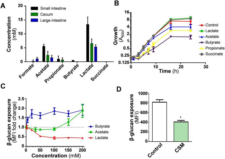
The immunogenicity of Candida albicans cells is influenced by changes in the exposure of microbe-associated molecular patterns (MAMPs) on the fungal cell surface. Previously, the degree of exposure on the C. albicans cell surface of the immunoinflammatory MAMP β-(1,3)-glucan was shown to correlate inversely with colonisation levels in the gastrointestinal (GI) tract. This is important because life-threatening systemic candidiasis in critically ill patients often arises from translocation of C. albicans strains present in the patient's GI tract. Therefore, using a murine model, we have examined the impact of gut-related factors upon β-glucan exposure and colonisation levels in the GI tract. The degree of β-glucan exposure was examined by imaging flow cytometry of C. albicans cells taken directly from GI compartments, and compared with colonisation levels. Fungal β-glucan exposure was lower in the cecum than the small intestine, and fungal burdens were correspondingly higher in the cecum. This inverse correlation did not hold for the large intestine. The gut fermentation acid, lactate, triggers β-glucan masking in vitro, leading to attenuated anti-Candida immune responses. Additional fermentation acids are present in the GI tract, including acetate, propionate, and butyrate. We show that these acids also influence β-glucan exposure on C. albicans cells in vitro and, like lactate, they influence β-glucan exposure via Gpr1/Gpa2-mediated signalling. Significantly, C. albicans gprgpa2Δ cells displayed elevated β-glucan exposure in the large intestine and a corresponding decrease in fungal burden, consistent with the idea that Gpr1/Gpa2-mediated β-glucan masking influences colonisation of this GI compartment. Finally, extracts from the murine gut and culture supernatants from the mannan grazing gut anaerobe Bacteroides thetaiotaomicron promote β-glucan exposure at the C. albicans cell surface. Therefore, the local microbiota influences β-glucan exposure levels directly (via mannan grazing) and indirectly (via fermentation acids), whilst β-glucan masking appears to promote C. albicans colonisation of the murine large intestine.

Keywords: Candida albicans; Cell wall; Fungal immunogenicity; Gut colonisation; β-Glucan exposure.


Conflict of interest statement

The authors declare that they have no known competing financial interests or personal relationships that could have appeared to influence the work reported in this paper.

Figures

None
Graphical abstract
Fig. 1
Fig. 1
C. albicans cells isolated from murine gut compartments display different levels of β-glucan exposure. Mice were pre-treated with antibiotics and then colonised with C. albicans SC5314 by oral gavage. After four days the mice were sacrificed, and the contents of their small intestine, cecum and large intestine taken for analysis. (A) Fungal burdens were quantified by measuring CFUs per gram of tissue. (B) The β-glucan exposure of C. albicans cells from each gut compartment was determined by imaging flow cytometry after staining with Fc-dectin-1 and anti-human IgG conjugated to Alexafluor 488. Median fluorescence intensities (MFIs) for each C. albicans population are presented: means and standard deviations for n = 3 mice. Statistical analyses were performed using the Mann-Whitney U test using GraphPad Prism 5: * p ≤ 0.05; ** p ≤ 0.01. (C) Two events (from n = 10,000) from the imaging flow cytometry are shown for each gut compartment: BF1, bright field 1; CH2, AF488 (β-glucan exposure); CH6, side scatter; CH7, Hoechst 33,342 (DNA); BF2, bright field 2; CH10, ConA-AF647 (mannan exposure). (D) Magnifications of the DIC and fluorescence images for representative C. albicans cells from each gut compartment are presented.
Fig. 2
Fig. 2
Bacterial fermentation acids in murine gut compartments and their impact on β-glucan exposure by C. albicans. (A) Fermentation acid concentrations were measured in the small intestine, cecum and large intestine of mice (n = 3) colonised for four days with C. albicans SC3154. (B) Impact of lactate (55 mM), acetate (83 mM), butyrate (56 mM), propionate (67 mM) and succinate (43 mM) upon the growth of C. albicans in GYNB at 30 °C. (C) Dose-dependent effects of lactate, acetate and butyrate upon β-glucan exposure levels for C. albicans SC5314 during growth in vitro in in GYNB at 30 °C. Cells were stained with Fc-dectin-1 (Fig. 1), their MFIs measured using a BD Fortessa flow cytometer, and β-glucan exposure levels expressed relative to the controls lacking SCFA. (D) The impact of colon simulating medium (CSM) on C. albicans β-glucan exposure, compared to control cells growing in GYNB. For all experiments, means and standard deviations are from three independent experiments (or n = 3 mice), and statistical analyses were performed using the Mann-Whitney U test using Prism 5: * p ≤ 0.05.
Fig. 3
Fig. 3
Impact of Gpr1/Gpa2 signalling on SCFA-mediated changes to C. albicans β-glucan exposure in vitro and in vivo. (A) Structural relationships between gut SCFAs. (B) The effects of SCFAs upon β-glucan exposure in C. albicans wild type control cells (Ca372) and the gprgpa2Δ double mutant (NM23). C. albicans were grown in vitro in GYNB at 30 °C and transferred to fresh GYNB either lacking or containing the SCFA for five hours. The cells were then stained with Fc-dectin-1 (Fig. 1), their MFIs measured using a BD Fortessa flow cytometer, and β-glucan exposure levels expressed relative to the control lacking SCFA: 55 mM lactate, 83 mM acetate, 56 mM butyrate, 67 mM propionate, and 43 mM succinate. (C) Mice (n = 3) were colonised with C. albicans wild type cells (Ca372) or the gprgpa2Δ double mutant (NM23). After four days the mice were sacrificed, and fungal burdens quantified (CFUs) in the small intestine, cecum and large intestine. (D) The levels of β-glucan exposure on C. albicans cells from the same gut compartments were measured by Fc-dectin-1 staining and imaging flow cytometry (Fig. 1). (E) Two events (from n = 10,000) from the imaging flow cytometry in Fig. 3D are shown for C. albicans wild type and gprgpa2Δ cells from each gut compartment: BF1, bright field 1; CH2, AF488 (β-glucan exposure); CH6, side scatter; CH7, Hoechst 33,342 (DNA); BF2, bright field 2; CH10, ConA-AF647 (mannan exposure). For all experiments, means and standard deviations are from three independent experiments (or n = 3 mice), and statistical analyses were performed using the Mann-Whitney U test using Prism 5: * p ≤ 0.05.
Fig. 4
Fig. 4
Impact of Wor1 on C. albicans β-glucan exposure and XOG1 expression levels in the gut. (A) Mice (n = 3) were colonised with C. albicans wild type (SC5314) or wor1Δ cells (CAY189), sacrificed after four days, and fungal burdens (CFUs) quantified in the small intestine, cecum and large intestine. (B) β-glucan exposure on C. albicans cells from the same gut compartments by imaging flow cytometry (Fig. 1). Means and standard deviations are from n = 3 mice, and statistical analyses were performed using the Mann-Whitney U test using Prism 5: * p ≤ 0.05. (C) RNA was prepared from the contents of the small intestine, cecum and large intestine, and from control C. albicans cells grown at 30 °C in GYNB. The levels of each target mRNA in these preparations were quantified by qRT-PCR relative to the internal ACT1 mRNA control, and then normalised against the transcript level in the control C. albicans cells grown in vitro. Means and standard deviations are from three independent experiments. The data were analysed statistically using one-way analysis of variance (ANOVA) using Prism 5: * p ≤ 0.05; ** p ≤ 0.01.
Fig. 5
Fig. 5
Effects of gut extracts upon β-glucan exposure and the C. albicans cell wall. (A) Antibiotic treated mice (n = 3) were sacrificed after four days, and soluble extracts prepared from their intestinal contents. A portion of these extracts was boiled to inactivate enzymes that might be present. In parallel, C. albicans SC5314 cells were pre-grown in GYNB at 30 °C, exposed to the colonic extracts, and the levels of β-glucan exposure on these cells at 0 and 6 h quantified by Fc-dectin-1 staining and imaging flow cytometry: Control untreated C. albicans cells, white; CE, cells treated with colonic extract, dark green; CE-boiled, cells treated with boiled colonic extract, pale green. Means and standard deviations are from three independent experiments, and statistical analyses were performed using the Mann-Whitney U test using Prism 5: ** p ≤ 0.01. (B) Images of representative cells from these assays by DIC and fluorescence microscopy. (C) Transmission electron microscopy (TEM) images of the cell walls of C. albicans cells incubated for 6 h with soluble extracts from the small intestine, cecum or large intestine of mice, and control C. albicans cells grown in vitro in GYNB at 30 °C. (For interpretation of the references to colour in this figure legend, the reader is referred to the web version of this article.). (For interpretation of the references to colour in this figure legend, the reader is referred to the web version of this article.)
Fig. 6
Fig. 6
Effects of culture supernatants from gut anaerobes on β-glucan exposure by, and immunogenicity of, C. albicans cells. (A) C. albicans SC5314 cells were grown in GYNB, fixed in thimerosal, and incubated with M2GSC growth medium only (control) or with culture supernatants from B. thetaiotaomicron B5482, B. adolescentis L2-32 or C. eutactus ART55/1 grown in M2GSC. Cells were then stained with Fc-dectin-1 (for β-glucan) and Concanavalin A (for mannan), and the levels of exposure of both MAMPs quantified using a BD Fortessa flow cytometer. Means and standard deviations for the MFIs from three independent experiments are presented, and the data analysed using the Mann-Whitney U test with Prism 5: * p ≤ 0.05; ** p ≤ 0.01. (B) Images of representative cells from these assays by DIC and fluorescence microscopy: β-glucan, green; mannan, red. (C) Fixed C. albicans cells from the 48 h timepoint were then incubated for 24 h with PBMCs (5 yeast to 1 PBMC) and TNF-α, IL-6 and IL-10 levels quantified. Each data point represents one sample of two from four different individuals. Means are presented, and the statistical analysis were performed using ANOVA with the Bonferroni post-hoc test: *, p ≤ 0.05; **, p ≤ 0.01; ***, p ≤ 0.001. (For interpretation of the references to colour in this figure legend, the reader is referred to the web version of this article.). (For interpretation of the references to colour in this figure legend, the reader is referred to the web version of this article.)

References

    1. Albenberg L., Esipova T.V., Judge C.P., Bittinger K., Chen J., Laughlin A., et al. Correlation between intraluminal oxygen gradient and radial partitioning of intestinal microbiota. Gastroenterology. 2014;147(5) 1055–1063.e8. - PMC - PubMed
    1. Alessi A.M., Gray V., Farquharson F.M., Flores-López A., Shaw S., Stead D., et al. β-Glucan is a major growth substrate for human gut bacteria related to Coprococcus eutactus. Environ. Microbiol. 2020;22(6):2150–2164. - PubMed
    1. Ballou E.R., Avelar G.M., Childers D.S., Mackie J., Bain J.M., Wagener J., et al. Lactate signalling regulates fungal beta-glucan masking and immune evasion. Nat. Microbiol. 2016;2:16238. - PMC - PubMed
    1. Barcenilla A., Pryde S.E., Martin J.C., Duncan S.H., Stewart C.S., Henderson C., et al. Phylogenetic relationships of butyrate-producing bacteria from the human gut. Appl. Environ. Microbiol. 2000;66(4):1654–1661. - PMC - PubMed
    1. Belenguer A., Duncan S.H., Calder A.G., Holtrop G., Louis P., Lobley G.E., et al. Two routes of metabolic cross-feeding between Bifidobacterium adolescentis and butyrate-producing anaerobes from the human gut. Appl. Environ. Microbiol. 2006;72(5):3593–3599. - PMC - PubMed

LinkOut - more resources