

Ontogenetic growth in the crania of Exaeretodon argentinus (Synapsida: Cynodontia) captures a dietary shift
- PMID: 36299507
- PMCID: PMC9590418
- DOI: 10.7717/peerj.14196
Ontogenetic growth in the crania of Exaeretodon argentinus (Synapsida: Cynodontia) captures a dietary shift
Abstract
Background: An ontogenetic niche shift in vertebrates is a common occurrence where ecology shifts with morphological changes throughout growth. How ecology shifts over a vertebrate's lifetime is often reconstructed in extant species-by combining observational and skeletal data from growth series of the same species-because interactions between organisms and their environment can be observed directly. However, reconstructing shifts using extinct vertebrates is difficult and requires well-sampled growth series, specimens with relatively complete preservation, and easily observable skeletal traits associated with ecologies suspected to change throughout growth, such as diet.
Methods: To reconstruct ecological changes throughout the growth of a stem-mammal, we describe changes associated with dietary ecology in a growth series of crania of the large-bodied (∼2 m in length) and herbivorous form, Exaeretodon argentinus (Cynodontia: Traversodontidae) from the Late Triassic Ischigualasto Formation, San Juan, Argentina. Nearly all specimens were deformed by taphonomic processes, so we reconstructed allometric slope using a generalized linear mixed effects model with distortion as a random effect.
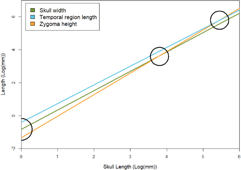
Results: Under a mixed effects model, we find that throughout growth, E. argentinus reduced the relative length of the palate, postcanine series, orbits, and basicranium, and expanded the relative length of the temporal region and the height of the zygomatic arch. The allometric relationship between the zygomatic arch and temporal region with the total length of the skull approximate the rate of growth for feeding musculature. Based on a higher allometric slope, the zygoma height is growing relatively faster than the length of the temporal region. The higher rate of change in the zygoma may suggest that smaller individuals had a crushing-dominated feeding style that transitioned into a chewing-dominated feeding style in larger individuals, suggesting a dietary shift from possible faunivory to a more plant-dominated diet. Dietary differentiation throughout development is further supported by an increase in sutural complexity and a shift in the orientation of microwear anisotropy between small and large individuals of E. argentinus. A developmental transition in the feeding ecology of E. argentinus is reflective of the reconstructed dietary transition across Gomphodontia, wherein the earliest-diverging species are inferred as omnivorous and the well-nested traversodontids are inferred as herbivorous, potentially suggesting that faunivory in immature individuals of the herbivorous Traversodontidae may be plesiomorphic for the clade.
Keywords: Allometry; Crania; Cyndont; Dietary ecology; Ecological differentiation; Exaeretodon; Ischigualasto formation; Ontogeny; Traversodontidae; Triassic.
©2022 Wynd et al.
Conflict of interest statement
The authors declare there are no competing interests.
Figures


Similar articles
-
Ontogeny of a Brazilian Late Triassic Traversodontid (Cynodontia, Cynognathia): Anatomical and Paleoecological Implications.J Morphol. 2025 Apr;286(4):e70047. doi: 10.1002/jmor.70047. J Morphol. 2025. PMID: 40249030 Free PMC article.
-
Evolution of postcanine complexity in Gomphodontia (Therapsida: Cynodontia).Anat Rec (Hoboken). 2024 Apr;307(4):1613-1633. doi: 10.1002/ar.25386. Epub 2024 Jan 28. Anat Rec (Hoboken). 2024. PMID: 38282465
-
Masticatory jaw movement of Exaeretodon argentinus (Therapsida: Cynodontia) inferred from its dental microwear.PLoS One. 2017 Nov 29;12(11):e0188023. doi: 10.1371/journal.pone.0188023. eCollection 2017. PLoS One. 2017. PMID: 29186178 Free PMC article.
-
Old fossil findings in the Upper Triassic rocks of southern Brazil improve diversity of traversodontid cynodonts (Therapsida, Cynodontia).Anat Rec (Hoboken). 2024 Apr;307(4):1474-1514. doi: 10.1002/ar.25244. Epub 2023 May 29. Anat Rec (Hoboken). 2024. PMID: 37246488
-
Including Distorted Specimens in Allometric Studies: Linear Mixed Models Account for Deformation.Integr Org Biol. 2021 May 18;3(1):obab017. doi: 10.1093/iob/obab017. eCollection 2021. Integr Org Biol. 2021. PMID: 34377943 Free PMC article.
Cited by
-
Ontogeny of a Brazilian Late Triassic Traversodontid (Cynodontia, Cynognathia): Anatomical and Paleoecological Implications.J Morphol. 2025 Apr;286(4):e70047. doi: 10.1002/jmor.70047. J Morphol. 2025. PMID: 40249030 Free PMC article.
-
Functional reorganisation of the cranial skeleton during the cynodont-mammaliaform transition.Commun Biol. 2023 Apr 12;6(1):367. doi: 10.1038/s42003-023-04742-0. Commun Biol. 2023. PMID: 37046052 Free PMC article.
References
-
- Abdala F, Giannini NP. Chiniquodontid cynodonts: systematic and morphometric considerations. Palaeontology. 2002;45:1151–1170. doi: 10.1111/1475-4983.00280. - DOI
-
- Abdala F, Malabarba MC. Enamel microstructure in Exaeretodon, a Late Triassic south american traversodontid (Therapsida: Cynodontia) Revista Brasileira de Paleontologia. 2007;10:71–78. doi: 10.4072/rbp.2007.2.01. - DOI
-
- Adams RA. Wing ontogeny, shifting niche dimensions, and adaptive landscapes. In: Adams RA, Pedersen SC, editors. Ontogeny, functional ecology, and evolution of bats. Cambridge University Press; Cambridge: 2000. pp. 275–315.
Publication types
MeSH terms
LinkOut - more resources
Full Text Sources
