

Isolation, characterization and anti-UVB irradiation activity of an extracellular polysaccharide produced by Lacticaseibacillus rhamnosus VHPriobi O17
- PMID: 36299523
- PMCID: PMC9589185
- DOI: 10.1016/j.heliyon.2022.e11125
Isolation, characterization and anti-UVB irradiation activity of an extracellular polysaccharide produced by Lacticaseibacillus rhamnosus VHPriobi O17
Abstract
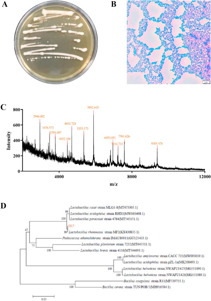
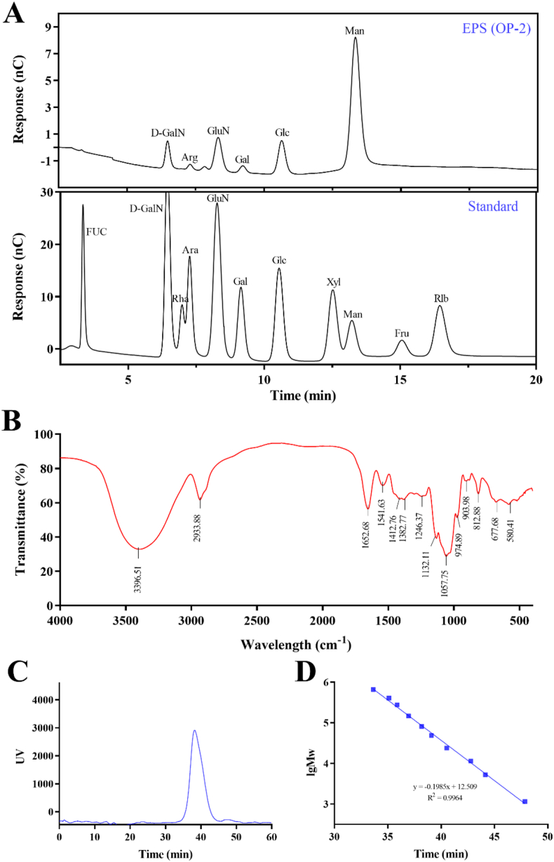
The purpose of this study was to isolate exopolysaccharides (EPS) from lactic acid bacteria (LAB) and evaluate EPS anti-UVB viability. Lacticaseibacillus rhamnosus VHPriobi O17 with high EPS production was screened from 34 strains of LAB. The EPS (OP-2) produced by L. rhamnosus VHPriobi O17 was purified by alcohol precipitation and DEAE-μSphere anion exchange chromatography. By ion chromatography, FT-IR spectrum and gel column chromatography, EPS (OP-2) was a novel Man-like polysaccharide with the weight-averaged molecular of 84.2 kDa. The EPS (OP-2) can effectively alleviate HaCaT cells apoptosis and overproduction of reactive oxygen species (ROS) induced by UVB. The results also showed that it inhibited the release of pro-inflammatory cytokines (IL-1α, IL-6 and IL-8); and suppressed the phosphorylation cascade of JNK and p38 MAPK to reduce the expression level of active-caspase3, ultimately prevented cell apoptosis. Thus, the EPS produced by L. rhamnosus VHPriobi O17 have the potential to be used for human anti-UVB irradiation.
Keywords: Anti-UVB; Exopolysaccharides; HaCaT cells; Lacticaseibacillus rhamnosus.
© 2022 The Author(s).
Conflict of interest statement
The authors declare no conflict of interest.
Figures


References
-
- Verschooten L., Claerhout S., Van Laethemii A., Agostinis P., Garmyn M. Invited review new strategies of photoprotection. Photochem. Photobiol. 2006;82:1016–1023. - PubMed
-
- Kammeyer A., Luiten R.M. Oxidation events and skin aging. Ageing Res. Rev. 2015;21:16–29. - PubMed
-
- Liu W., Wang F., Li C., Otkur W., Hayashi T., Mizuno K., Hattori S., Fujisaki H., Onodera S., Ikejima T. Silibinin treatment protects human skin cells from UVB injury through upregulation of estrogen receptors. J. Photochem. Photobiol. B Biol. 2021;216 - PubMed
-
- Xiao Z., Yang S., Chen J., Li C., Zhou C., Hong P., Sun S., Qian Z.J. Trehalose against UVB-induced skin photoaging by suppressing MMP expression and enhancing procollagen I synthesis in HaCaT cells. J. Funct.Foods. 2020;74
LinkOut - more resources
Full Text Sources
Research Materials
Miscellaneous
