Review
. 2022 Oct 8;25(11):105283.
doi: 10.1016/j.isci.2022.105283. eCollection 2022 Nov 18.

Role of non-cardiomyocytes in anticancer drug-induced cardiotoxicity: A systematic review

Affiliations
Review

Role of non-cardiomyocytes in anticancer drug-induced cardiotoxicity: A systematic review

Suifen Xie et al. iScience. .

Abstract

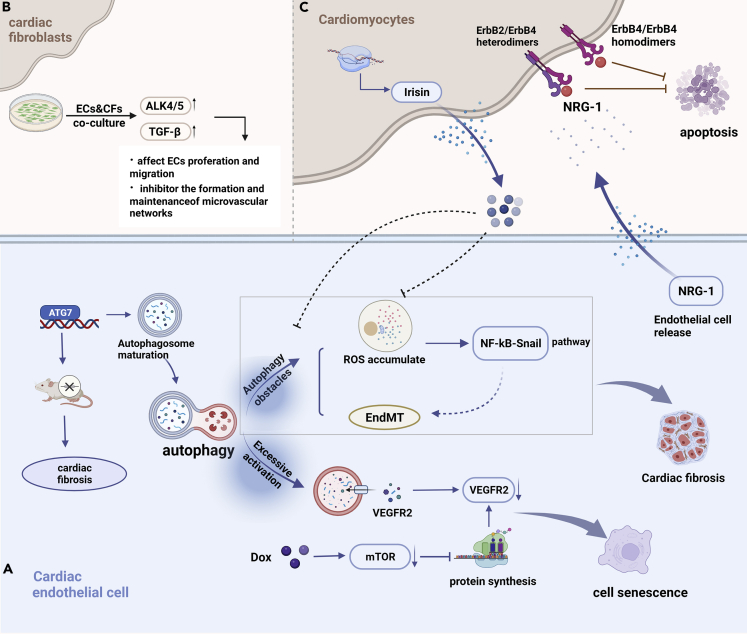
Cardiotoxicity induced by anticancer drugs interferes with the continuation of optimal treatment, inducing life-threatening risks or leading to long-term morbidity. The heart is a complex pluricellular organ comprised of cardiomyocytes and non-cardiomyocytes. Although the study of these cell populations has been often focusing on cardiomyocytes, the contributions of non-cardiomyocytes to development and disease are increasingly being appreciated as both dynamic and essential. This review summarized the role of non-cardiomyocytes in anticancer drug-induced cardiotoxicity, including the mechanism of direct damage to resident non-cardiomyocytes, cardiomyocytes injury caused by paracrine modality, myocardial inflammation induced by transient cell populations and the protective agents that focused on non-cardiomyocytes.

Keywords: Cardiovascular medicine; cancer; toxicity assessment.


Conflict of interest statement

The authors declare that they have no competing interests.

Figures

None
Graphical abstract
Figure 1
Figure 1
The regulation of anticancer drug-induced apoptosis and cardiac fibrosis in cardiac fibroblast Blunt-ended lines indicate inhibition while arrows indicate promotion; Apoptotic mechanism is on the left (blue) and fibrosis mechanism is on the right (pink). Both the MRP1-MRP1-GSH/GSSC pathway and increased NOX1 induced ROS accumulation and mitochondrial damage. Damaged mitochondria are well known as DAMPs that activate the innate immune system. The release of DAMPs from damaged mitochondria increases and is recognized by Toll-like receptor 9, which induces myocardial fibrosis. MMPs family (mmp1, mmp2, mmp7, and so forth) can induce myocardial fibrosis by activating the PAR-1 receptor or mmp1-PI3K-Akt pathway. Among them, Akt binds to the FasL receptor in cardiomyocytes after activation, thereby inducing apoptosis of cardiomyocytes, and rosmarinic acid can effectively alleviate it. Abbreviations: PAR-1, protease-activated receptor-1; MMPs, matrix metalloproteinase; Dox, doxorubicin; MRP1, motility-related protein 1; GSH, reduced glutathione; GSSG, oxidized glutathione; NOX1, NADPH oxidase 1; ROS, reactive oxygen species; TM5541, an inhibitor of plasminogen activator inhibitor-1; DAMPs, damage-associated molecular patterns; LZDO, liguzinediol; RA, rosmarinic acid; FasL, Fas ligand; Akt, protein kinase B; PI3K, phosphoinositide3-kinase; P53, tumor suppressor protein; Gβ5, G Protein subunit beta 5; NK-1R, neurokinin-1 receptor; TLR4, Toll-like receptor 4; TLR9, toll-like receptor 9; OND2088, TLR-9 antagonist; IL-1β, interleukin 1 beta; IL-6, interleukin 6; TIMP-1, tissue inhibitor of metalloproteinase-1; TGF-β, transforming growth factor β; P substance, a neuropeptide.
Figure 2
Figure 2
Signaling regulation in endothelial cells (A) the regulation mechanism of cardiac endothelial cell senescence and cardiac fibrosis. Dox-induced overactivation of autophagy and inhibition of mTOR degrades VEGFR2 in a short time and decline continuously, promoting endothelial cell senescence. EndMT is a phenotypic transformation process in which endothelial cells gradually lose their own characteristics and acquire interstitial characteristics. The activation of NF- κ B-nial pathway induced by ROS may be a potential way of EndMT, and autophagy obstacle is the cause of ROS accumulation. (B) The interaction between endothelial cells and fibroblasts inhibits endothelial cell proliferation. Irisin is a novel hormone-like myokine that ameliorates autophagy disorders and EndMT by regulating UCP2. NRG-1 is an active cardiac growth factor released by endothelial cells, which plays an anti-apoptotic effect by combining with ErbB2/ErbB4 heterodimer on the surface of cardiomyocytes. (C) the interaction between endothelial cells and cardiomyocytes induces apoptosis and myocardial fibrosis. Abbreviations: Endothelial cells, endothelial cells; Cardiac fibroblasts, cardiac fibroblasts; ALK4/5, a type of TGF-β receptor; ErbB2/ErbB4, a member of the ErbB family of receptor tyrosine kinases; NRG-1, neuromodulatory protein-1; ATG7, autophagy-related gene 7; NF-κB, nuclear factor kappa light chain enhancer of activated B cells; EndMT, endothelial-to-mesenchymal transition; VEGFR2, vascular endothelial growth factor receptor 2; mTOR, mammalian target of rapamycin.
Figure 3
Figure 3
Immune cell-induced cardiotoxicity (A) Schematic diagram of myocardial inflammation induced by immune checkpoint inhibitor therapy. PD-1 and CTLA-4 are two critical immune checkpoints that regulate T cell immune responses. Immune checkpoint inhibitors can inhibit negative immune regulation, resulting in adverse immune events such as myocarditis. The dominant manifestation of myocarditis is T lymphocyte and macrophage infiltration. (B) Myocarditis caused by T lymphocyte infiltration. Transdifferentiation of CD4+ cells into Th1 induced by TNF-γ promotes cardiac injury and activation of CD8+ cells, which further mediates myocardial fibrosis and cardiac inflammation. (C) Myocarditis caused by Macrophage infiltration. It was found that macrophages are able to attain either M1 or M2 phenotype, with M1 acting as a pro-inflammatory but M2 promoting tissue repair. PD-1 does not cause direct damage to cardiomyocytes, but induces damage through the transfer of miR-34a-5p to cardiomyocytes through macrophage-derived exosomes. Abbreviations: PD-1, programmed cell death protein-1; CTLA-4, cytotoxic T lymphocyte-associated antigen-4; TCR, T-cell receptor; CMs, cardiomyocytes.

References

    1. Ahmed S.H., Clark L.L., Pennington W.R., Webb C.S., Bonnema D.D., Leonardi A.H., Mcclure C.D., Spinale F.G., Zile M.R. Matrix metalloproteinases/tissue inhibitors of metalloproteinases: relationship between changes in proteolytic determinants of matrix composition and structural, functional, and clinical manifestations of hypertensive heart disease. Circulation. 2006;113:2089–2096. - PubMed
    1. Anderson R., Theron A.J., Nel J.G., Durandt C., Cholo M.C., Feldman C., Tintinger G.R. Clofazimine, but not isoniazid or rifampicin, augments platelet activation in vitro. Front. Pharmacol. 2018;9:1335. - PMC - PubMed
    1. Ando J., Yamamoto K. Flow detection and calcium signalling in vascular endothelial cells. Cardiovasc. Res. 2013;99:260–268. - PubMed
    1. Banerjee I., Fuseler J.W., Price R.L., Borg T.K., Baudino T.A. Determination of cell types and numbers during cardiac development in the neonatal and adult rat and mouse. Am. J. Physiol. Heart Circ. Physiol. 2007;293:H1883–H1891. - PubMed
    1. Brutsaert D.L. Cardiac endothelial-myocardial signaling: its role in cardiac growth, contractile performance, and rhythmicity. Physiol. Rev. 2003;83:59–115. - PubMed

LinkOut - more resources