

Derivation, validation and assessment of a novel nomogram-based risk assessment model for venous thromboembolism in hospitalized patients with lung cancer: A retrospective case control study
- PMID: 36300098
- PMCID: PMC9589115
- DOI: 10.3389/fonc.2022.988287
Derivation, validation and assessment of a novel nomogram-based risk assessment model for venous thromboembolism in hospitalized patients with lung cancer: A retrospective case control study
Abstract
Purpose: This study aimed to develop and validate a specific risk-stratification nomogram model for the prediction of venous thromboembolism(VTE) in hospitalized patients with lung cancer using readily obtainable demographic, clinical and therapeutic characteristics, thus guiding the individualized decision-making on thromboprophylaxis on the basis of VTE risk levels.
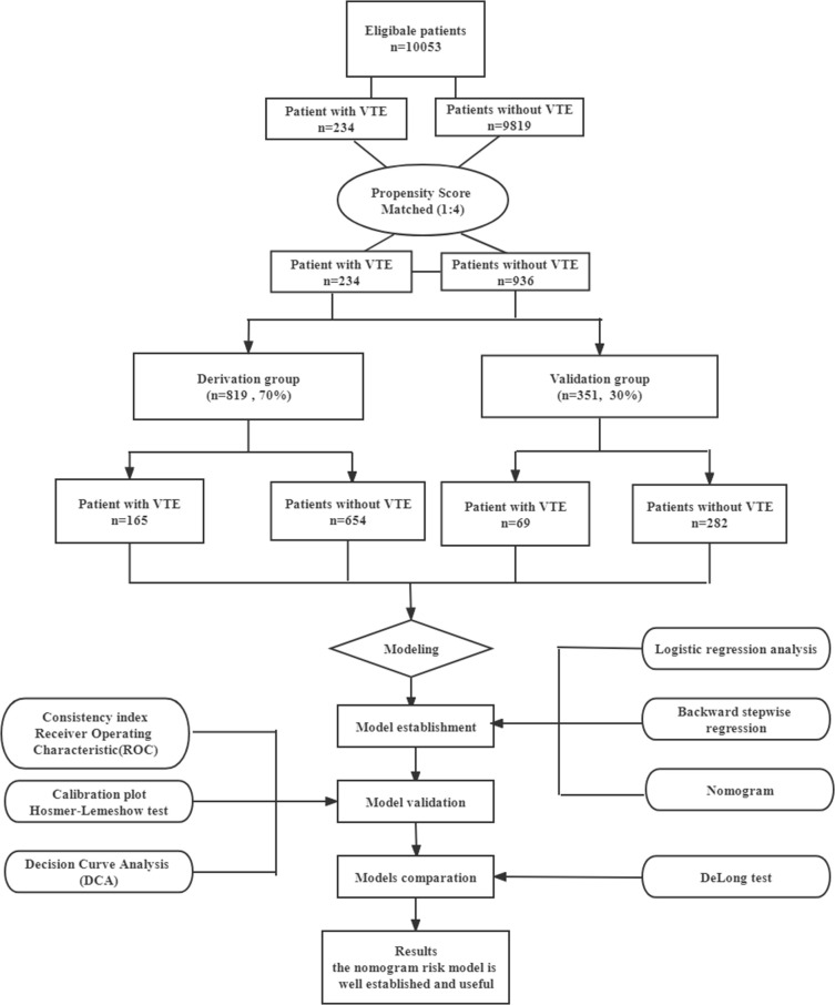
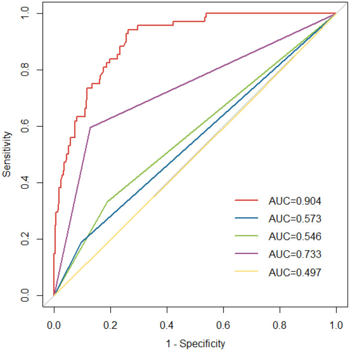
Methods: We performed a retrospective case-control study among newly diagnosed lung cancer patients hospitalized between January 2016 and December 2021. Included in the cohort were 234 patients who developed PTE and 936 non-VTE patients. The patients were randomly divided into the derivation group (70%, 165 VTE patients and 654 non-VTE patients) and the validation group (30%, 69 VTE patients and 282 non-VTE patients). Cut off values were established using a Youden´s Index. Univariate and multivariate regression analyses were used to determine independent risk factors associated with VTE. Variance Inflation Factor(VIF) was used for collinearity diagnosis of the covariates in the model. The model was validated by the consistency index (C-index), receiver operating characteristic curves(ROC) and the calibration plot with the Hosmer-Lemeshow goodness-of-fit test. The clinical utility of the model was assessed through decision curve analysis(DCA). Further, the comparison of nomogram model with current models(Khorana, Caprini, Padua and COMPASS-CAT) was performed by comparing ROC curves using the DeLong's test.
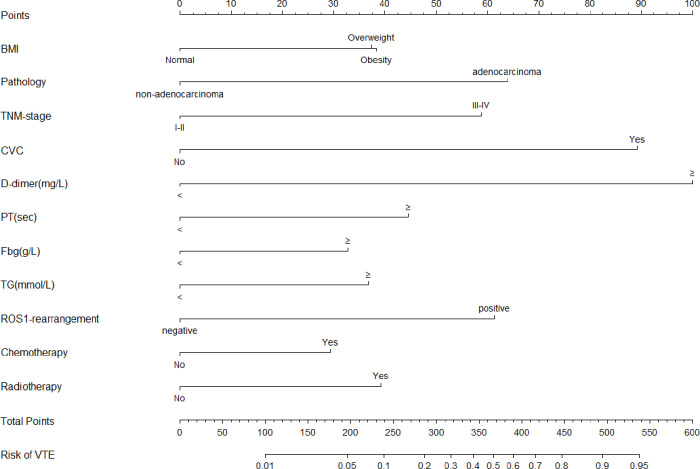
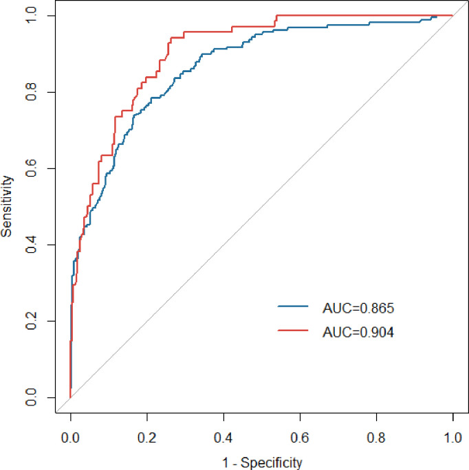
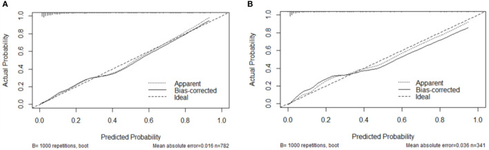
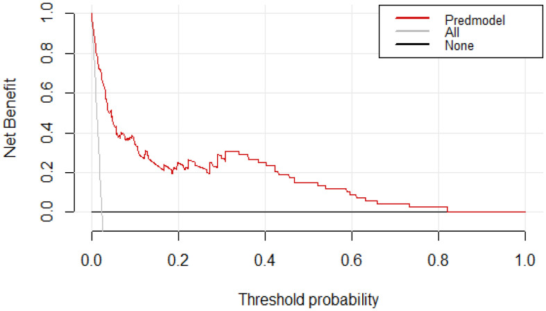
Results: The predictive nomogram modle comprised eleven variables: overweight(24-28) defined by body mass index (BMI): [odds ratio (OR): 1.90, 95% confidence interval (CI): 1.19-3.07], adenocarcinoma(OR:3.00, 95% CI: 1.88-4.87), stageIII-IV(OR:2.75, 95%CI: 1.58-4.96), Central venous catheters(CVCs) (OR:4.64, 95%CI: 2.86-7.62), D-dimer levels≥2.06mg/L(OR:5.58, 95%CI:3.54-8.94), PT levels≥11.45sec(OR:2.15, 95% CI:1.32-3.54), Fbg levels≥3.33 g/L(OR:1.76, 95%CI:1.12-2.78), TG levels≥1.37mmol/L (OR:1.88, 95%CI:1.19-2.99), ROS1 rearrangement(OR:2.87, 95%CI:1.74-4.75), chemotherapy history(OR:1.66, 95%CI:1.01-2.70) and radiotherapy history(OR:1.96, 95%CI:1.17-3.29). Collinearity analysis with demonstrated no collinearity among the variables. The resulting model showed good predictive performance in the derivation group (AUC 0.865, 95% CI: 0.832-0.897) and in the validation group(AUC 0.904,95%CI:0.869-0.939). The calibration curve and DCA showed that the risk-stratification nomogram had good consistency and clinical utility. Futher, the area under the ROC curve for the specific VTE risk-stratification nomogram model (0.904; 95% CI:0.869-0.939) was significantly higher than those of the KRS, Caprini, Padua and COMPASS-CAT models(Z=12.087, 11.851, 9.442, 5.340, all P<0.001, respectively).
Conclusion: A high-performance nomogram model incorporated available clinical parameters, genetic and therapeutic factors was established, which can accurately predict the risk of VTE in hospitalized patients with lung cancer and to guide individualized decision-making on thromboprophylaxis. Notably, the novel nomogram model was significantly more effective than the existing well-accepted models in routine clinical practice in stratifying the risk of VTE in those patients. Future community-based prospective studies and studies from multiple clinical centers are required for external validation.
Keywords: lung cancer; nomogram model; risk assessment; thromboprophylaxis; venous thromboembolism.
Copyright © 2022 Li, Tian, Niu, He, Cao, Zhang, Kaiweisierkezi and Luo.
Conflict of interest statement
The authors declare that the research was conducted in the absence of any commercial or financial relationships that could be construed as a potential conflict of interest.
Figures
References
LinkOut - more resources
Full Text Sources
Miscellaneous