. 2022 Dec;50(6):140.
doi: 10.3892/ijmm.2022.5196. Epub 2022 Oct 27.

Therapeutic function of a novel rat induced pluripotent stem cell line in a 6‑OHDA‑induced rat model of Parkinson's disease

Affiliations

Therapeutic function of a novel rat induced pluripotent stem cell line in a 6‑OHDA‑induced rat model of Parkinson's disease

Jiajia Xu et al. Int J Mol Med. 2022 Dec.

Abstract

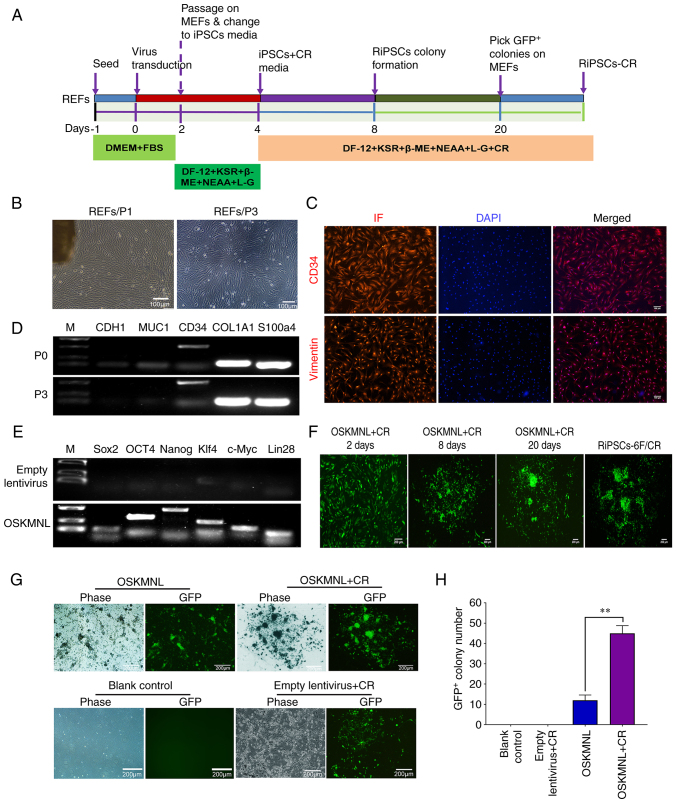
Parkinson's disease (PD) is a progressive neurodegenerative movement disorder of the central nervous system that results from the loss of dopaminergic (DA) nigral neurons. Induced pluripotent stem cells (iPSCs) have shown potential for cell transplantation treatment of neurodegenerative disorders. In the present study, the small molecules CHIR99021 and RepSox (CR) significantly facilitated reprogramming and enhanced the efficiency of GFP+/iPS‑like colonies [rat iPSCs induced by OCT3/4, Sox2, Klf4, c‑Myc, Nanog and Lin28 + CR (RiPSCs‑6F/CR)] generation by ~4.0‑fold during lentivirus‑mediated reprogramming of six transcription factors in rat embryonic fibroblasts. The generation of iPSCs was detected by reverse transcription‑PCR, immunofluorescence and western blot analysis. Subsequently, RiPSCs‑6F/CR were stereotactically transplanted into the right medial forebrain bundle (MFB) of 6‑hydroxydopamine‑lesioned rats with PD. The transplanted RiPSCs‑6F/CR survived and functioned in the MFB of rats with PD for ≥20 weeks, and significantly improved functional restoration from their PD‑related behavioral defects. Furthermore, the grafted RiPSCs‑6F/CR could migrate and differentiate into various neurocytes in vivo, including γ aminobutyric acid‑ergic, DA neurons and glial cells. In conclusion, the present study confirmed that RiPSCs‑6F/CR induced by small molecules could be used as potential donor material for neural grafting to remodel basal ganglia circuitry in neurodegenerative diseases.

Keywords: Parkinson's disease; cell reprogramming; neural transplantation; rat induced pluripotent stem cells; small molecules.


Conflict of interest statement

The authors declare that they have no competing interests.

Figures

Figure 1
Figure 1
Induction and characterization of RiPSCs-6F/CR generated from REFs. (A) Schematic representation of the OSKMNL-CR transduction reprogramming protocol. (B) Morphology of REFs at P1 and P3. Fibroblast-like cells could be seen migrating from the tissue pieces in the P1 image. (C) REFs expressed fibroblast markers CD34 and Vimentin, as observed by IF staining (scale bars, 100 µm). (D) Expression of fibroblast- and epithelial-specific marker genes was detected by reverse transcription-PCR. (E) All OSKMNL genes were overexpressed 3 days after infection of REFs with OSKMNL-expressing lentiviral particles. According to the size of the PCR amplification fragment, the upper bands were determined as the target bands. The bands below are primer dimers of PCR amplification, which may be attributed to the low annealing temperature and high primer concentration. (F) Cell morphological changes throughout the induction process. (G) GFP+/iPS-like colonies were induced by OSKMNL and OSKMNL + CR; CR markedly promoted the efficiency of GFP+/iPS-like colonies generation. (H) Statistical analysis of the number of GFP+ clones. **P<0.01 vs. OSKMNL+CR. RiPSCs-6F/CR, rat iPSCs induced by OSKMNL + CR; REFs, rat embryonic fibroblasts; MEFs, mouse embryonic fibroblasts; iPSCs, induced pluripotent stem cells; OSKMNL, OCT3/4, Sox2, Klf4, c-Myc, Nanog and Lin28; DF-12, DMEM/F-12; DMEM, Dulbecco's modified Eagle's medium; KSR, KnockOut Serum Replacement; β-ME, β-mercaptoethanol; NEAA, non-essential amino acid; L-G, L-glutamine; CR, CHIR99021 and RepSox; P, passage; IF, immunofluorescence.
Figure 2
Figure 2
Biological characterization of RiPSCs-6F/CR. (A) AP-positive dense cell colonies of RiPSCs-6F/CR. (B) Chromosome G-banding analysis of RiPSCs-6F/CR. (C) RiPSCs-6F/CR expressed the pluripotency markers OCT4, Sox2 and Nanog, as observed by immunofluorescence (scale bars, 100 µm). (D) Expression of OCT4 and Sox2 in RiPSCs-6F/CR was detected using flow cytometry. (E) Expression of six transcription factor genes was detected by reverse transcription PCR. (F) Expression of pluripotency markers was detected by western blotting. RiPSCs-6F/CR, rat induced pluripotent stem cells induced by OCT3/4, Sox2, Klf4, c-Myc, Nanog and Lin28 + CR; CR, CHIR99021 and RepSox; AP, alkaline phosphatase.
Figure 3
Figure 3
RiPSCs-6F/CR significantly ameliorate the behavioral deficits of a rat model of Parkinson's disease. (A) 6-Hydroxydopamine was stereotactically transplanted into the right medial forebrain bundle of each model rat at the labeled two coordinates. (B) Behavioral evaluation by sniff test, ipsilateral rotation and rotarod test. (C) Experimental rats were fixed on a stereotactic apparatus. (D) Transplantation of RiPSCs-6F/CR did not lead to tumor formation at 8 weeks after transplantation. (E) Heat maps of open-field assay. (F-H) Motor behavior evaluation in three groups at 8 and 24 weeks after transplantation. *P<0.05, **P<0.01 and ***P<0.001 vs. RiPSCs-6F/CR. RiPSCs-6F, rat induced pluripotent stem cells induced by OSKMNL; RiPSCs-6F/CR, rat induced pluripotent stem cells induced by OSKMNL + CR; OSKMNL, OCT3/4, Sox2, Klf4, c-Myc, Nanog and Lin28; CR, CHIR99021 and RepSox.
Figure 4
Figure 4
Effects of RiPSCs-6F/CR transplantation on the loss of TH+ dopaminergic neurons in the right medial forebrain bundle of rats with Parkinson's disease. (A) Representative images of whole brain scanning with hematoxylin and eosin staining in the healthy control, vehicle and RiPSCs-6F/CR groups 12 weeks after transplantation. (B) TH intensity was detected by TH-3,3'-diaminobenzidine staining of the whole brain in three groups, and was analyzed using CaseViewer. The boxed areas are shown at higher magnification on the right side of the image. (C) Number of TH+ cells in the injured areas of the three groups. *P<0.05 by one-way ANOVA. RiPSCs-6F/CR, rat induced pluripotent stem cells induced by OCT3/4, Sox2, Klf4, c-Myc, Nanog and Lin28 + CR; CR, CHIR99021 and RepSox; TH, tyrosine hydroxylase.
Figure 5
Figure 5
Survival and migration of RiPSCs-6F/CR in the right medial forebrain bundle of rats with Parkinson's disease. (A) RiPSCs-6F/CR formed a distinct graft area and migrated 2.3 mm from the graft site 20 weeks after transplantation. (B) Grafted RiPSCs-6F/CR differentiated into various functional neurons in vivo, which could express TH, GABA and TUJ1 20 weeks after transplantation. (C) Numerous grafted cells were positive for PSD95 staining. The transplanted RiPSCs-6F/CR were labeled by white arrows. RiPSCs-6F/CR, rat induced pluripotent stem cells induced by OCT3/4, Sox2, Klf4, c-Myc, Nanog and Lin28 + CR; CR, CHIR99021 and RepSox; TH, tyrosine hydroxylase; GABA, γ aminobutyric acid; TUJ1, βIII tubulin; PSD95, postsynaptic density protein 95.
Figure 6
Figure 6
Differentiation and integration of RiPSCs-6F/CR cells in the brain of rats with Parkinson's disease. (A) RiPSCs-6F/CR could differentiate into GFAP+ glial cells. Grafted RiPSCs-6F/CR differentiated into neural stem cells or neural precursor cells, and expressed the neural stem markers (B) Sox2, (C) Nestin and (D) PAX6 20 weeks after transplantation. (E) Approximately 52% of the transplanted RiPSCs-6F/CR cells expressed the synapse marker SYN 20 weeks after transplantation. RiPSCs-6F/CR, rat induced pluripotent stem cells induced by OCT3/4, Sox2, Klf4, c-Myc, Nanog and Lin28 + CR; CR, CHIR99021 and RepSox; GFAP, glial fibrillary acidic protein; PAX6, paired box protein 6; SYN, synaptophysin.

Similar articles

Cited by

References

    1. Guo Y, Guan Y, Zhu H, Sun T, Wang Y, Huang Y, Ma C, Emery R, Guan W, Wang C, Liu C. Therapeutic function of iPSCs-derived primitive neuroepithelial cells in a rat model of Parkinson's disease. Neurochem Int. 2022;155:105324. doi: 10.1016/j.neuint.2022.105324. - DOI - PubMed
    1. Tang Y, Meng L, Wan CM, Liu ZH, Liao WH, Yan XX, Wang XY, Tang BS, Guo JF. Identifying the presence of Parkinson's disease using low-frequency fluctuations in BOLD signals. Neurosci Lett. 2017;645:1–6. doi: 10.1016/j.neulet.2017.02.056. - DOI - PubMed
    1. Parmar M, Grealish S, Henchcliffe C. The future of stem cell therapies for Parkinson disease. Nat Rev Neurosci. 2020;21:103–115. doi: 10.1038/s41583-019-0257-7. - DOI - PubMed
    1. Armstrong MJ, Okun MS. Diagnosis and treatment of Parkinson disease: A review. JAMA. 2020;323:548–560. doi: 10.1001/jama.2019.22360. - DOI - PubMed
    1. Blesa J, Przedborski S. Parkinson's disease: Animal models and dopaminergic cell vulnerability. Front Neuroanat. 2014;8:155. doi: 10.3389/fnana.2014.00155. - DOI - PMC - PubMed