Risk Factors for Perioperative Brain Lesions in Infants With Congenital Heart Disease: A European Collaboration
- PMID: 36300371
- PMCID: PMC9698124
- DOI: 10.1161/STROKEAHA.122.039492
Risk Factors for Perioperative Brain Lesions in Infants With Congenital Heart Disease: A European Collaboration
Abstract
Background: Infants with congenital heart disease are at risk of brain injury and impaired neurodevelopment. The aim was to investigate risk factors for perioperative brain lesions in infants with congenital heart disease.
Methods: Infants with transposition of the great arteries, single ventricle physiology, and left ventricular outflow tract and/or aortic arch obstruction undergoing cardiac surgery <6 weeks after birth from 3 European cohorts (Utrecht, Zurich, and London) were combined. Brain lesions were scored on preoperative (transposition of the great arteries N=104; single ventricle physiology N=35; and left ventricular outflow tract and/or aortic arch obstruction N=41) and postoperative (transposition of the great arteries N=88; single ventricle physiology N=28; and left ventricular outflow tract and/or aortic arch obstruction N=30) magnetic resonance imaging for risk factor analysis of arterial ischemic stroke, cerebral sinus venous thrombosis, and white matter injury.
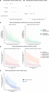
Results: Preoperatively, induced vaginal delivery (odds ratio [OR], 2.23 [95% CI, 1.06-4.70]) was associated with white matter injury and balloon atrial septostomy increased the risk of white matter injury (OR, 2.51 [95% CI, 1.23-5.20]) and arterial ischemic stroke (OR, 4.49 [95% CI, 1.20-21.49]). Postoperatively, younger postnatal age at surgery (OR, 1.18 [95% CI, 1.05-1.33]) and selective cerebral perfusion, particularly at ≤20 °C (OR, 13.46 [95% CI, 3.58-67.10]), were associated with new arterial ischemic stroke. Single ventricle physiology was associated with new white matter injury (OR, 2.88 [95% CI, 1.20-6.95]) and transposition of the great arteries with new cerebral sinus venous thrombosis (OR, 13.47 [95% CI, 2.28-95.66]). Delayed sternal closure (OR, 3.47 [95% CI, 1.08-13.06]) and lower intraoperative temperatures (OR, 1.22 [95% CI, 1.07-1.36]) also increased the risk of new cerebral sinus venous thrombosis.
Conclusions: Delivery planning and surgery timing may be modifiable risk factors that allow personalized treatment to minimize the risk of perioperative brain injury in severe congenital heart disease. Further research is needed to optimize cerebral perfusion techniques for neonatal surgery and to confirm the relationship between cerebral sinus venous thrombosis and perioperative risk factors.
Keywords: heart diseases; ischemic stroke; magnetic resonance imaging; pedatrics; risk factors; venous thrombosis; white matter.
Figures


References
-
- Hoffman JIE, Kaplan S. The incidence of congenital heart disease. J Am Coll Cardiol. 2002;39:1890–1900. doi: 10.1016/s0735-1097(02)01886-7 - PubMed
-
- Marino BS, Lipkin PH, Newburger JW, Peacock G, Gerdes M, Gaynor JW, Mussatto KA, Uzark K, Goldberg CS, Johnson WHJ., et al. . Neurodevelopmental outcomes in children with congenital heart disease: evaluation and management: a scientific statement from the American Heart Association. Circulation. 2012;126:1143–1172. doi: 10.1161/CIR.0b013e318265ee8a - PubMed
-
- Feldmann M, Bataillard C, Ehrler M, Ullrich C, Knirsch W, Gosteli-Peter MA, Held U, Latal B. Cognitive and executive function in congenital heart disease: a meta-analysis. Pediatrics. 2021;148:70–95. doi: 10.1542/peds.2021-050875 - PubMed
-
- Miller SP, McQuillen PS, Hamrick S, Xu D, Glidden DV, Charlton N, Karl T, Azakie A, Ferriero DM, Barkovich AJ., et al. . Abnormal brain development in newborns with congenital heart disease. N Engl J Med. 2007;357:1928–1938. doi: 10.1056/NEJMoa067393 - PubMed
Publication types
MeSH terms
Grants and funding
LinkOut - more resources
Full Text Sources
Medical

