

Radial nerve entrapment after fracture of the supracondylar humerus: a rare case of a 6-year-old
- PMID: 36300558
- PMCID: PMC9682549
- DOI: 10.5152/j.aott.2022.22062
Radial nerve entrapment after fracture of the supracondylar humerus: a rare case of a 6-year-old
Erratum in
-
Erratum.Acta Orthop Traumatol Turc. 2022 Nov;56(6):421. doi: 10.5152/j.aott.2022.23001. Acta Orthop Traumatol Turc. 2022. PMID: 36441051 Free PMC article. No abstract available.
Abstract
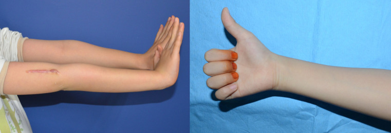
Supracondylar fracture of the humerus is one of the most common fractures seen in children, and posteromedial displacement of the distal fragment in extension-type supracondylar humerus fractures can cause injury to the radial nerve. A 6-year old girl who presented with symptoms of radial nerve injury after a supracondylar fracture of the right humerus with complete posteromedial displacement of the distal fragment (Gartland type III) underwent surgery where closed reduction and percutaneous pinning was performed. The patient was routinely followed up and at 6 months postoperatively no neurological improvement was seen. Exploratory surgery revealed complete discontinuation of the radial nerve at the fracture site and entrapment of the nerve stumps in healed bone callus. A gap of 2 cm was observed between nerve stumps, and sural nerve cable grafting was performed with good results. If neurological symptoms do not improve over time, appropriate differential diagnosis and, if necessary, exploratory surgery should be considered. Despite limited reports and their conflicting outcomes, sural nerve cable grafting could be a useful option to bridge the gap of discontinued nerve injury. Level of Evidence: Level IV, Case Report.
Figures


Similar articles
-
[Radiographic and clinical assessment of supracondylar humeral fractures resulted from sports in children].Zhonghua Yi Xue Za Zhi. 2017 Jan 17;97(3):208-211. doi: 10.3760/cma.j.issn.0376-2491.2017.03.010. Zhonghua Yi Xue Za Zhi. 2017. PMID: 28162172 Chinese.
-
Radial nerve palsies associated with paediatric supracondylar humeral fractures: a caution in the interpretation of neurophysiological studies.J Pediatr Orthop B. 2020 Mar;29(2):126-132. doi: 10.1097/BPB.0000000000000680. J Pediatr Orthop B. 2020. PMID: 31567895
-
Gartland Type III Pediatric Supracondylar Humerus Fracture with Radial Nerve Laceration: A Case Report.JBJS Case Connect. 2023 Mar 3;13(1). doi: 10.2106/JBJS.CC.22.00658. eCollection 2023 Jan 1. JBJS Case Connect. 2023. PMID: 36870051
-
Compartment syndrome of the upper arm after closed reduction and percutaneous pinning of a supracondylar humerus fracture.J Pediatr Orthop. 2014 Mar;34(2):e1-4. doi: 10.1097/BPO.0b013e3182933c69. J Pediatr Orthop. 2014. PMID: 23774207 Review.
-
Radial Nerve Palsy After Humeral Shaft Fractures: The Case for Early Exploration and a New Classification to Guide Treatment and Prognosis.Hand Clin. 2018 Feb;34(1):105-112. doi: 10.1016/j.hcl.2017.09.011. Hand Clin. 2018. PMID: 29169591 Review.
Cited by
-
Intramedullary nailing using K-wires for high-energy distal humeral metaphyseal-diaphyseal fractures accompanying radial nerve palsy in a 2-year-old toddler: A case report.Medicine (Baltimore). 2025 Jul 25;104(30):e43322. doi: 10.1097/MD.0000000000043322. Medicine (Baltimore). 2025. PMID: 40725908 Free PMC article.
-
Isolated Radial Nerve Palsy Following Supracondylar Humerus Fracture in a Pediatric Patient: A Case Report.Cureus. 2024 Oct 30;16(10):e72677. doi: 10.7759/cureus.72677. eCollection 2024 Oct. Cureus. 2024. PMID: 39618659 Free PMC article.
References
Publication types
MeSH terms
LinkOut - more resources
Full Text Sources
Medical
Miscellaneous
