. 2022 Dec 8;7(23):e162392.
doi: 10.1172/jci.insight.162392.

Intestinal epithelial BLT1 promotes mucosal repair

Affiliations

Intestinal epithelial BLT1 promotes mucosal repair

Shusaku Hayashi et al. JCI Insight. .

Abstract

Acute and chronic intestinal inflammation is associated with epithelial damage, resulting in mucosal wounds in the forms of erosions and ulcers in the intestinal tract. Intestinal epithelial cells (IECs) and immune cells in the wound milieu secrete cytokines and lipid mediators to influence repair. Leukotriene B4 (LTB4), a lipid chemokine, binds to its receptor BLT1 and promotes migration of immune cells to sites of active inflammation; however, a role for intestinal epithelial BLT1 during mucosal wound repair is not known. Here we report that BLT1 was expressed in IECs both in vitro and in vivo, where it functioned as a receptor not only for LTB4 but also for another ligand, resolvin E1. Intestinal epithelial BLT1 expression was increased when epithelial cells were exposed to an inflammatory microenvironment. Using human and murine primary colonic epithelial cells, we reveal that the LTB4/BLT1 pathway promoted epithelial migration and proliferation leading to accelerated epithelial wound repair. Furthermore, in vivo intestinal wound repair experiments in BLT1-deficient mice and bone marrow chimeras demonstrated an important contribution of epithelial BLT1 during colonic mucosal wound repair. Taken together, our findings show a potentially novel prorepair in IEC mechanism mediated by BLT1 signaling.

Keywords: Gastroenterology; Inflammation; Inflammatory bowel disease.


Figures

Figure 1
Figure 1. BLT1 functions as a major epithelial receptor for RvE1.
(A) RNAscope staining for LTB4R and CMKLR1 mRNA expression in frozen sections from colonic tissue of humans. (B) RNAscope staining for Ltb4r1 and Cmklr1 mRNA expression in frozen sections from colonic tissue of mice. Scale bars: 50 μm. (C) qPCR analysis of the expression of CMKLR1 and LTB4R mRNA in the SKCO-15, T84, and human 2D colonoids. The data are presented as the mean ± SEM. Cq, quantification cycle (measured as cycles). (D) Effect of BLT1 antagonist on the prorepair activity of RvE1 in the scratch wound assay using human primary IECs. After scratch wound was produced, IECs were incubated with RvE1 (100 nM) for 24 hours. BLT1 (CP105,696; 1 μM) or CMKLR1 (α-NETA; 10 μM) antagonist was applied 30 minutes before RvE1 treatment. Quantification of wound repair at 24 hours after wounding is shown. The data are presented as the mean ± SEM. Statistical analysis was performed using 1-way ANOVA followed by post hoc Welch’s t test with Bonferroni’s correction. *P < 0.05, **P < 0.01, compared with RvE1.
Figure 2
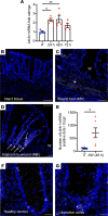
Figure 2. Epithelial BLT1 is upregulated in response to colonic mucosal injury.
(A) The changes in the expression of Ltb4r1 mRNA in 3 mm punch biopsies of intact colonic tissues and colonic mucosal wounds on different days after injury. The data are presented as the mean ± SEM of 4–5 mice. Statistical analysis was performed using 1-way ANOVA followed by post hoc Welch’s t test with Bonferroni’s correction. *P < 0.05, **P < 0.01, compared with intact tissue (IT). (BD) RNAscope staining for Blt1 mRNA in frozen sections from intact tissues and wounded colonic tissues 2 days after injury. Arrows indicate upregulation of Ltb4r1 expression in the crypts next to the wound. W, wound. Scale bar is 50 μm. (E) The number of Ltb4r1 mRNA–positive dots in the crypt of intact colonic tissues and colonic mucosal wounds (adjacent to wound) 2 days after injury is shown. The data are presented as the mean ± SEM of 6 mice. Statistical analysis was performed using an unpaired (2-tailed) t test with Welch’s correction. *P < 0.05, compared with IT. AW, adjacent to wound. (F and G) RNAscope staining for LTB4R mRNA expression in frozen sections from healthy controls and patients with ulcerative colitis.
Figure 3
Figure 3. BLT1 regulates intestinal epithelial wound repair.
(A) Effect of LTB4 in the scratch wound assay using SKCO-15 cells. The data are presented as the mean ± SEM. Statistical analysis was performed using 1-way ANOVA followed by post hoc Welch’s t test with Bonferroni’s correction. *P < 0.05; **P < 0.01. (B and C) Effect of LTB4 in the scratch wound assay using human primary colonic epithelial monolayers. (B) Representative phase-contrast images at 0 and 24 hours after wounding are shown. Scale bar is 100 μm. (C) Quantification of change over time in wound repair is shown. The data are presented as the mean ± SEM. Statistical analysis was performed using 2-way ANOVA followed by post hoc Welch’s t test with Bonferroni’s correction. **P < 0.01; ***P < 0.001; ****P < 0.0001, compared with vehicle. P < 0.05; ††P < 0.01: †††P < 0.001; ††††P < 0.0001, compared with LTB4. (D and E) Effect of LTB4 in the scratch wound assay using primary epithelial monolayers. (D) Representative phase-contrast images at 0 and 24 hours after wounding are shown. Scale bar is 100 μm. (E) Quantification of change over time in wound repair is shown. The data are presented as the mean ± SEM. Statistical analysis was performed using 2-way ANOVA followed by post hoc Welch’s t test with Bonferroni’s correction. *P < 0.05; ****P < 0.0001, compared with WT (vehicle). ††P < 0.01; ††††P < 0.0001, compared with Ltb4r1–/– (vehicle). (F) qPCR analysis of the changes in the expression of LTB4R mRNA in the human 2D cultured colonoid stimulated with IFN-γ (10 ng/mL) and TNF-α (10 ng/mL). The data are presented as the mean ± SEM. Statistical analysis was performed using an unpaired (2-tailed) t test with Welch’s correction. *P < 0.05. NT, nontreated. (G) Effect of IFN-γ (100 ng/mL) and TNF-α (100 ng/mL) on the prorepair activity of low-dose LTB4 (1 nM) in the scratch wound assay using SKCO-15 cells. The data are presented as the mean ± SEM. Statistical analysis was performed using 1-way ANOVA followed by post hoc Welch’s t test with Bonferroni’s correction. **P < 0.01; ***P < 0.001; ****P < 0.0001.
Figure 4
Figure 4. BLT1 activation promotes migration and proliferation of IECs.
(AD) Migration analysis by DiPer. (A) Plot at the origin graph of 20 cells. (B) Mean square displacement of 20 cells. The data are presented as the mean ± SEM. Statistical analysis was performed using 2-way ANOVA followed by post hoc Welch’s t test with Bonferroni’s correction. **P < 0.01, ****P < 0.0001, compared with WT (vehicle). ††P < 0.01, †††P < 0.001, ††††P < 0.0001, compared with Ltb4r1–/– (vehicle). (C) Velocity autocorrelation was measured on at least 20 cells. Statistical analysis was performed using 2-way ANOVA followed by post hoc Welch’s t test with Bonferroni’s correction. **P < 0.01, ***P < 0.001, ****P < 0.0001, compared with WT (vehicle). ††P < 0.01, compared with Ltb4r1–/– (vehicle). (D) Average cell speed was calculated on 20 cells. The data are presented as the mean ± SEM. Statistical analysis was performed using 1-way ANOVA followed by post hoc Welch’s t test with Bonferroni’s correction. ***P < 0.001, ****P < 0.0001. (E) Immunoblotting was performed on lysates from scratch-wounded IEC monolayers treated with LTB4 (100 nM) or vehicle. Levels of phosphorylated SRC (p-SRC) (Y416) and p-FAK (Y397, Y925) were compared with total Src, FAK, and GAPDH to assess activation. Numbers on the left represent kDa. (F and G) EdU incorporation analysis in murine 3D cultured colonoids stimulated with LTB4 (10 nM) for 24 hours. (F and G) Effect of BLT1 antagonist. Pictures show representative images of EdU-incorporated (shown in green) colonoids. Blue, nuclei. Scale bar is 10 μm. The data are presented as the mean ± SEM. Statistical analysis was performed using 1-way ANOVA followed by post hoc Welch’s t test with Bonferroni’s correction. *P < 0.05, **P < 0.01.
Figure 5
Figure 5. Role of BLT1 in intestinal mucosal wound repair in vivo.
(A) In vivo intestinal mucosal wound repair in Ltb4r1–/– mice. Utilizing a miniature video endoscope and biopsy scissors, 5 wounds were created in the dorsal aspect of the colonic mucosa of anesthetized mice. Digital images of wound surface area at 1 and 3 days after wounding are shown (left). Points represent the mean value within all wounds from individual mice (right). The data are presented as the mean ± SEM of 9 to 10 mice. Statistical analysis was performed using an unpaired (2-tailed) t test with Welch’s correction. ****P < 0.0001. (B and C) In vivo intestinal mucosal wound repair in BM chimeric mice. (B) Illustration of BM chimera experiment. (C) Digital images of wound surface area at 1 and 3 days after wounding are shown (left). Points represent the mean value within all wounds from individual mice (right). The data are presented as the mean ± SEM of 5 mice. Statistical analysis was performed using 1-way ANOVA followed by post hoc Welch’s t test with Bonferroni’s correction. ***P < 0.001, ****P < 0.0001.

Similar articles

Cited by

References

    1. Brazil JC, et al. Innate immune cell-epithelial crosstalk during wound repair. J Clin Invest. 2019;129(8):2983–2993. doi: 10.1172/JCI124618. - DOI - PMC - PubMed
    1. Quiros M, Nusrat A. Saving problematic mucosae: SPMs in intestinal mucosal inflammation and repair. Trends Mol Med. 2019;25(2):124–135. doi: 10.1016/j.molmed.2018.12.004. - DOI - PubMed
    1. Serhan CN, Levy BD. Resolvins in inflammation: emergence of the pro-resolving superfamily of mediators. J Clin Invest. 2018;128(7):2657–2669. doi: 10.1172/JCI97943. - DOI - PMC - PubMed
    1. Quiros M, et al. Resolvin E1 is a pro-repair molecule that promotes intestinal epithelial wound healing. Proc Natl Acad Sci U S A. 2020;117(17):9477–9482. doi: 10.1073/pnas.1921335117. - DOI - PMC - PubMed
    1. Arita M, et al. Resolvin E1 selectively interacts with leukotriene B4 receptor BLT1 and ChemR23 to regulate inflammation. J Immunol. 2007;178(6):3912–3917. doi: 10.4049/jimmunol.178.6.3912. - DOI - PubMed

Publication types