. 2023 Jan 3;133(1):e154684.
doi: 10.1172/JCI154684.

Mitochondrial dysfunction reactivates α-fetoprotein expression that drives copper-dependent immunosuppression in mitochondrial disease models

Affiliations

Mitochondrial dysfunction reactivates α-fetoprotein expression that drives copper-dependent immunosuppression in mitochondrial disease models

Kimberly A Jett et al. J Clin Invest. .

Abstract

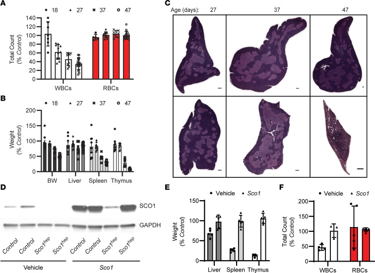
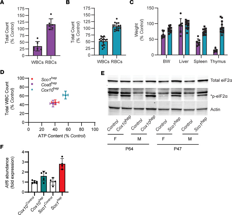
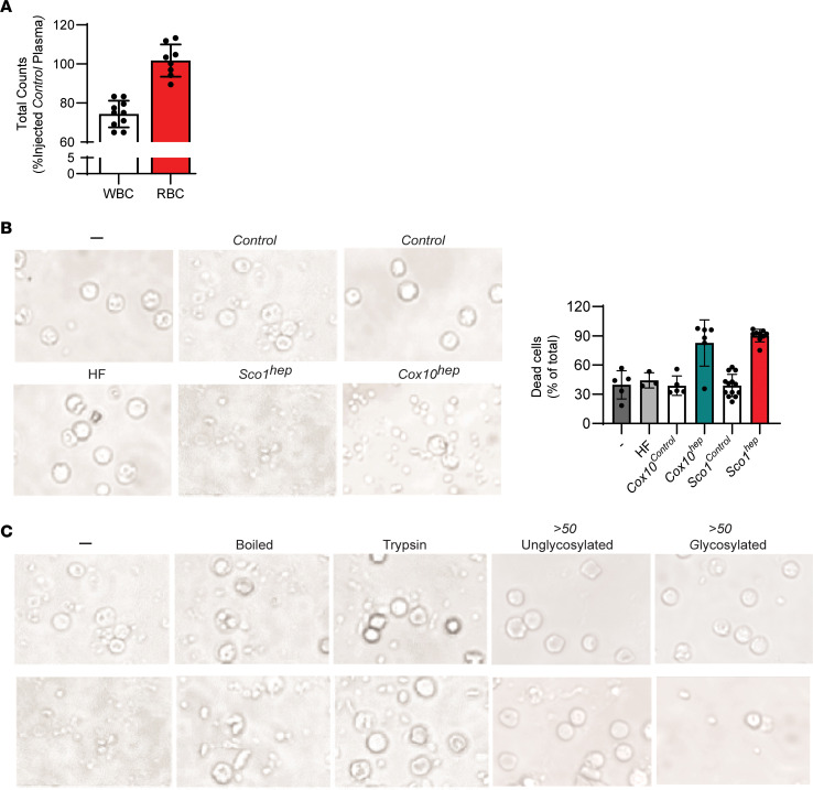
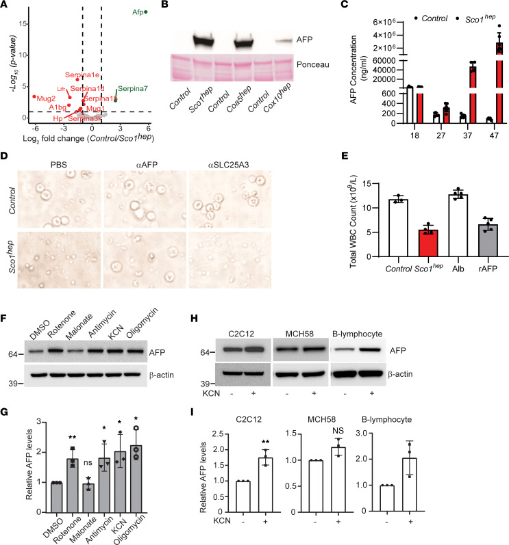
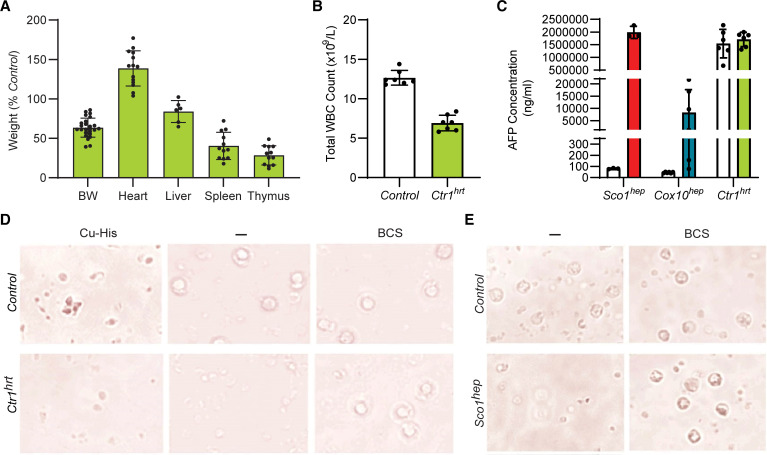
Signaling circuits crucial to systemic physiology are widespread, yet uncovering their molecular underpinnings remains a barrier to understanding the etiology of many metabolic disorders. Here, we identified a copper-linked signaling circuit activated by disruption of mitochondrial function in the murine liver or heart that resulted in atrophy of the spleen and thymus and caused a peripheral white blood cell deficiency. We demonstrated that the leukopenia was caused by α-fetoprotein, which required copper and the cell surface receptor CCR5 to promote white blood cell death. We further showed that α-fetoprotein expression was upregulated in several cell types upon inhibition of oxidative phosphorylation. Collectively, our data argue that α-fetoprotein may be secreted by bioenergetically stressed tissue to suppress the immune system, an effect that may explain the recurrent or chronic infections that are observed in a subset of mitochondrial diseases or in other disorders with secondary mitochondrial dysfunction.

Keywords: Metabolism; Mitochondria.


Conflict of interest statement

Conflict of interest: The authors have declared that no conflict of interest exists.

Figures

Figure 1
Figure 1. Ablation of Sco1 expression in hepatocytes results in an unexpected reduction in circulating white blood cell counts and atrophy of the thymus and spleen.
(A) Progressive leukopenia in Sco1hep mice (2-way ANOVA with Bonferroni’s post hoc test, n = 5–20; P < 0.01) and (B) disproportionate reduction in the wet weights of the Sco1hep spleen and thymus at P37 (n = 8; P < 0.01) and P47 (n = 13; P < 0.001); 2-tailed Student’s t test. (C) Selective thinning of the Sco1hep thymic cortex. Top row: Control. Bottom row: Sco1hep. Scale bars: 2 mm and 800 μm (P47 Sco1hep thymus). (D) Adenoviral restoration of SCO1 expression in the liver leads to (E) rescue of splenic and thymic atrophy (n = 5) and (F) normalization of WBC counts (n = 3–5). Mice were injected on P21 via cardiac puncture with vehicle or helper-dependent adenovirus harboring a Sco1 cDNA under the control of a liver-specific promoter and harvested at P47. Control, wild-type littermates; BW, body weight; WBC and RBC, white and red blood cells, respectively.
Figure 2
Figure 2. The reduction in peripheral WBC counts in hepatocytes is positively correlated with the bioenergetic deficit in the liver.
(A) Coa5hep (n = 6; P < 0.001) and (B) Cox10hep (n = 15–16; P < 0.01) mice also exhibit a significant leukopenia. (C) Coa5hep mice (purple bars) have a disproportionate reduction in the wet weight of the spleen (n = 6; P < 0.01) and thymus (n = 5; P < 0.001), while Cox10hep mice (blue bars) exhibit significant yet milder atrophy of the spleen (n = 14; P < 0.05). Significance was assessed using a 2-tailed Student’s t test (AC). (D) Total WBC counts are positively correlated with liver ATP content in all 3 hep mouse models (linear regression R2 = 0.99; P = 0.001). P47 Sco1hep and P64 Cox10hep livers have higher levels of (E) the phosphorylated form of eIF2α, an ISR marker, and (F) the Atf6 transcript, a marker of ER stress (1-way ANOVA with Tukey’s post hoc test, n = 4–6; P < 0.005, Sco1hep model; P < 0.02, Cox10hep model).
Figure 3
Figure 3. A glycoprotein secreted by mice with altered mitochondrial function in the liver is responsible for the observed reduction in peripheral WBC counts.
(A) Total WBC counts are reduced in Control mice injected with Sco1hep plasma relative to those injected with Control plasma (n = 10; P = 0.02 by 2-tailed Student’s t test). (B) The viability of PBMCs isolated from Control mice is significantly reduced when they are cultured with Sco1hep or Cox10hep plasma. PBMC viability is unaffected by exposure to plasma from mice fed a high-fat (HF) diet. Bar graph on right summarizes the operator-blinded quantitation of dead cells per treatment group expressed as a percentage of the total number of cells (live and dead) per image (n = 3–13 per treatment group, P < 0.0001, both hep models versus PBMCs cultured in FBS alone [–], HF, or Control treatment). (C) PBMC viability is rescued if Sco1hep plasma is boiled or treated with trypsin. Fractionation based on size and the presence of a glycan revealed that the factor(s) that reduces PBMC viability is present in a >50 kDa glycoprotein fraction. Scale bars: 50 μm (B and C). The Control and Sco1hep images in panel B are identical to those denoted as “–” in C because they were part of the same experimental series.
Figure 4
Figure 4. AFP expression is increased in response to impaired organelle function and is directly responsible for the leukopenia observed in several mitochondrial disease models.
(A) AFP is significantly enriched in hep relative to Control plasma. Volcano plot with dotted lines indicating a 2-fold change and adjusted P-value significance threshold of 0.1. A green or red symbol indicates a protein whose abundance is significantly up- or downregulated, respectively. Gray symbols denote proteins whose abundance is not significantly different (NS) between the 2 groups. (B) AFP abundance is markedly upregulated in Sco1hep, Coa5hep, and Cox10hep plasma. Plasma was pooled (minimum of 2 males and 2 females per pool) and depleted of immunoglobulins and albumin prior to Western blotting. The Ponceau S–stained membrane indicates relative loading across lanes. (C) AFP progressively accumulates in Sco1hep plasma (n = 4–7; P27, P < 0.05; P37, P < 0.0001; P47, P < 0.0001). (D) PBMC viability is rescued by immunodepleting AFP from Sco1hep plasma. Culture media supplemented with PBS and plasma incubated with αSLC25A3, an antibody isotype control, served as internal controls. Original magnification, ×60 (same scale as in Figure 3, B and C; Figure 5, D and E; and Figure 6, C and D). (E) Control mice develop a leukopenia following serial injection with Sco1hep plasma (n = 3–4; P < 0.01) or 1 μg of recombinant AFP (rAFP) (n = 5; P < 0.01). Control mice injected with Control plasma or albumin served as internal controls. (F and G) Inhibition of the mitochondrial respiratory chain elevates AFP abundance in C2C12 myoblasts. (H and I) AFP levels increase in C2C12 myoblasts and human B lymphoblasts upon inhibition of COX. For panels FI, β-actin was used as a loading control and data are shown as mean ± SD (n = 3). *P < 0.05, **P < 0.005. Significance was assessed using a 2-tailed Student’s t test (C, E, G, and I). NS, not significant.
Figure 5
Figure 5. AFP requires copper to promote the death of peripheral WBCs.
(A) Ctr1hrt mice exhibit disproportionate atrophy of the spleen (n = 12; P < 0.01) and thymus (n = 12; P < 0.01) and have a (B) leukopenia (n = 7; P < 0.01). Significance was assessed using a 2-tailed Student’s t test (A and B). (C) Plasma AFP levels are comparable in P10 Control and Ctr1hrt plasma mice. Relative levels of AFP in Sco1hep, Cox10hep, and age-matched littermate Control plasma were quantified at the same time and are shown here for comparative purposes (n = 3–6). (D) PBMC viability is adversely affected by Ctr1hrt plasma, an effect that can be rescued by adding the copper chelator bathocuproine sulphonate (BCS) to the media. Viability of PBMCs is also reduced if copper-histidine (Cu-His) is added to media containing P10 Control plasma. (E) BCS also negates the negative effect of Sco1hep plasma on PBMC viability. Scale bars: 50 μm (D and E).
Figure 6
Figure 6. AFP promotes activation and apoptosis of both mouse and human WBCs via the cell surface receptor CCR5.
(A) PBMCs isolated from peripheral blood of Sco1hep mice have an increased number of cells that stain positive for the activation marker CD44 and the apoptotic marker annexin V when compared with PBMCs isolated from Controls (n = 6). (B) PBMCs isolated from Control mice that were cultured in media containing Sco1hep plasma are activated earlier and demonstrate a progressive increase in cell surface expression of the apoptotic marker annexin V relative to PBMCs cultured in Control plasma (n = 3). (C) Cells analyzed in B show blebbing (a sign of apoptosis) as early as 12–24 hours in culture and loss of cellularity after 72 hours in culture in response to culturing with Sco1hep but not Control plasma. (D) Human PBMC viability is also reduced in media containing Sco1hep plasma, and can be rescued with the CCR5 antagonist maraviroc. mPBMCs and hPBMCs, PBMCs of mouse and human origin, respectively (mPBMCs, n = 3; hPBMCs n = 2; each replicate contained a triplicate for each experimental condition). Scale bars: 50 μm (C and D).

Similar articles

Cited by

References

    1. Rajan A, Perrimon N. Drosophila as a model for interorgan communication: lessons from studies on energy homeostasis. Dev Cell. 2011;21(1):29–31. doi: 10.1016/j.devcel.2011.06.034. - DOI - PMC - PubMed
    1. Taylor RC, et al. Systemic stress signalling: understanding the cell non-autonomous control of proteostasis. Nat Rev Mol Cell Biol. 2014;15(3):211–217. doi: 10.1038/nrm3752. - DOI - PMC - PubMed
    1. Deng P, Haynes CM. The mitokine quest(ion) Cell Res. 2016;26(12):1265–1266. doi: 10.1038/cr.2016.138. - DOI - PMC - PubMed
    1. Schinzel R, Dillin A. Endocrine aspects of organelle stress—cell non-autonomous signaling of mitochondria and the ER. Curr Opin Cell Biol. 2015;33:102–110. doi: 10.1016/j.ceb.2015.01.006. - DOI - PubMed
    1. Droujinine IA, Perrimon N. Interorgan communication pathways in physiology: focus on drosophila. Annu Rev Genet. 2016;50:539–570. doi: 10.1146/annurev-genet-121415-122024. - DOI - PMC - PubMed

Publication types