Review
. 2023 Jul;12(7):399-427.
doi: 10.1089/wound.2022.0059. Epub 2022 Dec 13.

Recent Advances in Functional Wound Dressings

Affiliations
Review

Recent Advances in Functional Wound Dressings

Huy Quang Tran et al. Adv Wound Care (New Rochelle). 2023 Jul.

Abstract

Significance: Nowadays, the wound dressing is no longer limited to its primary wound protection ability. Hydrogel, sponge-like material, three dimensional-printed mesh, and nanofiber-based dressings with incorporation of functional components, such as nanomaterials, growth factors, enzymes, antimicrobial agents, and electronics, are able to not only prevent/treat infection but also accelerate the wound healing and monitor the wound-healing status. Recent Advances: The advances in nanotechnologies and materials science have paved the way to incorporate various functional components into the dressings, which can facilitate wound healing and monitor different biological parameters in the wound area. In this review, we mainly focus on the discussion of recently developed functional wound dressings. Critical Issues: Understanding the structure and composition of wound dressings is important to correlate their functions with the outcome of wound management. Future Directions: "All-in-one" dressings that integrate multiple functions (e.g., monitoring, antimicrobial, pain relief, immune modulation, and regeneration) could be effective for wound repair and regeneration.

Keywords: chronic wound; diabetic foot ulcer; hydrogels; nanofibers; sponges; wound dressings; wound healing.


Conflict of interest statement

No competing financial interests exist. The content of this article was entirely written by the authors listed. No ghost-writers were used to write this article.

Figures

None
Jingwei Xie, PhD
Figure 1.
Figure 1.
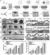
Schematic representation for different types of functional wound dressings for non-healing wound applications.
Figure 2.
Figure 2.
SF-based electrospun nanofibers loaded with antimicrobial VKC peptide displayed excellent antimicrobial property against gram-positive and gram-negative bacteria with high water uptake efficiency and cell-stimulatory capability in vitro, and they further facilitated re-epithelialization and neovascularization at the wound area in diabetic mouse models. (A) Representative preparation of VKC and VKC loaded SF electrospun nanofibers and their application in induced diabetic wounds. (B) Wound evolution after being treated with SF nanofibers with and without antimicrobial agent. (C) The wound closure rates at different time points of four groups. (D) Quantification of length of wound area at day 9. (E) Hematoxylin and eosin staining of four groups at different time points. Statistical significance (**p < 0.01, ***p < 0. 001). VKC, vitamin K3 carnosine peptide. SF, silk fibroin. Adapted from Agarwal et al. with permission.
Figure 3.
Figure 3.
Biphasic scaffolds featuring dissolvable microneedles rendered its ability for deep penetration into the structures of bacterial biofilms, facilitating the sustained release of therapeutic agents and improving its combinatory efficiency in biofilm eradication. (A, B) Schematic illustrating the fabrication of biphasic scaffolds. (A) Preparation of antimicrobial agents/F127-PCL core-sheath nanofiber mats. (B) Preparation of dissolvable PVP microneedles with antimicrobial agents and their assembly to nanofiber mats. (C–E) A representative SEM image of immobilized AgGaVan containing high-density PVP microneedle arrays and F127/AgGaVan-PCL core-sheath nanofiber mat serving as a substrate for a biphasic scaffold. (F) Long-term sustained release profiles of silver ions, vancomycin, and gallium ions in vitro. (G, H) The efficacy of biphasic scaffolds with different formulations against mixed-MRSA/P. aeruginosa biofilms ex vivo using different administration strategies. (G) Scaffolds containing a high density of microneedle arrays were changed three times within 108 h and (H) 144 h. Statistical significance (*p < 0.05). Adapted from Su et al. with permission. PCL, polycaprolactone; PVP, polyvinylpyrrolidone.
Figure 4.
Figure 4.
The Zn2+ incorporated PCP functional hydrogels possessed extraordinary mechanical properties with high flexibility and capability of self-healing, shaped remodeling; in addition, on the exposure to electrical stimulation, they performed a better wound-healing capacity in infected diabetic wounds compared with that of commercial product Hydrosorb® with more organized collagen fibers and granulation tissue formation at the wound site. (A) Schematic illustration for the preparation of Zn2+ incorporated conductive PCP hydrogels. (B) Self-healing capacity and stretchability of the PCPZ hydrogels. (C) The recovery of infected diabetic wounds being treated with commercial dressing Hydrosorb, PCPZ hydrogels with and without electrical stimulation. (D) Hematoxylin & eosin and Masson staining of restored tissues at the site of injuries. Adapted from Zhang et al. with permission. PCP, poly(vinyl alcohol)/chitosan/polypyrrole.
Figure 5.
Figure 5.
Rhein@OR-S hydrogels became unstable and dissociated under oxidative stress, releasing self-assembled Rhein at the wound site, which assisted the alleviation of excess reactive oxygen species. The hydrogels demonstrated a fast-healing property toward diabetic wounds with a closer wound gap in diabetic mouse after treatment. (A) Schematic presentation for the preparation of oxidation responsive Rhein@OR-S hydrogels and its mechanism under oxidative stress in a chronic wound environment. (B) Alterations of different hydrogels regarding their modulus strength and pore diameters on exposure to oxidative stress. (C) Progress of wound recovery in diabetic mouse models being treated with different hydrogels. (D) Hematoxylin–eosin staining of the tissues from the wound edges on day 14. Adapted from Zhao et al. with permission.
Figure 6.
Figure 6.
NS-rG@VEGF sponge scaffold facilitated the transmission of bioelectrical signals in the wound microenvironment and accelerated the restoration of the opened wound in the diabetic mouse model. Besides that, the synergistic effect of VEGF and rG under the endogenous ES stimulated not only the collagen deposition but also the enhanced amount of neovascularization and restored functional hair follicles (A) Schematic presentation for the preparation 3D NS-rG@VEGF sponge scaffold and its utilization for diabetic wound healing. (B) Wound evolution after being treated with different sponge compositions. (C) Representative Masson's staining of wounds treated with different sponge compositions. (D) The quantification for α-SMA density from different groups. (E) The number of restored hair follicles from different groups. Statistical significance (*p < 0.05, **p < 0.01). Adapted from Wang et al. with permission. NS nanofibrous sponge. rG reduced graphene oxide. α-SMA, α-smooth muscle actin; VEGF, vascular endothelial growth factor.
Figure 7.
Figure 7.
Three dimensional-printed SIS/MBG@Exos dressing promoted angiogenesis and enhanced the re-epithelialization process in diabetic wounds. (A) Schematic illustration for the preparation of cryogenic 3D printing SIS/MBG@Exos hydrogel scaffolds. (B) The process of wound remedy being treated with 3D-printed scaffolds. (C, D) Hematoxylin–eosin and Masson staining in different treated groups, respectively. (E) Immunofluorescence α-SMA-probing to present neovascularization of the diabetic wound. Adapted from Hu et al. with permission. SIS decellularized small intestinal submucosa. MBG, mesoporous bioactive glass. Exos, exosomes; 3D, three dimensional.
Figure 8.
Figure 8.
The incorporation of phenol red, Gox, and HRP into the hydrogel matrix of PCB enabled its capability to monitor the value of pH value and level of glucose in the wound area by measuring the intensity of RGB signals obtained from images taken from a smartphone device; in addition, the biosensor dressing maintained a moist environment to stimulate the wound-healing process in diabetic rats. (A) Representative illustration for the components and functionality of PCB hydrogel. (B) RGB images of as-prepared PCB hydrogel sensor with different glucose concentrations or pH values. (C) The evolution of wound healing in healthy and diabetic mice with different dressings. (D) Hematoxylin–eosin staining of the tissues from the wound edges on day 14. (E) Size of wound areas with different dressings from day 0 to day 14. (F) Quantification of the epithelial gaps on day 14. Statistical significance (*p < 0.05, **p < 0.01). Adapted from Zhu et al. with permission. GOx, glucose oxidase; HRP, horseradish peroxidase; PCB, polycarboxybetaine.

Similar articles

Cited by

References

    1. Sen CK. Human wounds and its burden: An updated compendium of estimates. Adv Wound Care 2019;2:39–48. - PMC - PubMed
    1. Tottoli EM, Dorati R, Genta I, et al. . Skin wound healing process and new emerging technologies for skin wound care and regeneration. Pharmaceutics 2020;12:e735. - PMC - PubMed
    1. Krishnan KA, Thomas S. Recent advances on herb-derived constituents-incorporated wound-dressing materials: a review. Polym Adv Technol 2019;30:823–838.
    1. Powers JG, Higham C, Broussard K, et al. . Wound healing and treating wounds: chronic wound care and management. J Am Acad Dermatol 2016;74:607–625. - PubMed
    1. FrykbergRobert G. Challenges in the treatment of chronic wounds. Adv Wound Care 2015;4:560–582. - PMC - PubMed

Publication types