Review
. 2023 Feb;38(4-6):338-351.
doi: 10.1089/ars.2022.0151.

Chicken or Egg? Mitochondrial Phospholipids and Oxidative Stress in Disuse-Induced Skeletal Muscle Atrophy

Affiliations
Review

Chicken or Egg? Mitochondrial Phospholipids and Oxidative Stress in Disuse-Induced Skeletal Muscle Atrophy

Edwin R Miranda et al. Antioxid Redox Signal. 2023 Feb.

Abstract

Significance: Accumulation of reactive oxygen species (ROS) is known to promote cellular damage in multiple cell types. In skeletal muscle, ROS has been implicated in disuse-induced muscle atrophy. However, the molecular origin and mechanism of how disuse promotes ROS and muscle dysfunction remains unclear. Recent Advances: Recently, we implicated membrane lipids of mitochondria to be a potential source of ROS to promote muscle atrophy. Critical Issues: In this review, we discuss evidence that changes in mitochondrial lipids represent a physiologically relevant process by which disuse promotes mitochondrial electron leak and oxidative stress. Future Directions: We further discuss lipid hydroperoxides as a potential downstream mediator of ROS to induce muscle atrophy. Antioxid. Redox Signal. 38, 338-351.

Keywords: ROS; disuse; lipid peroxidation; mitochondrial phospholipids; myopathy.


Conflict of interest statement

The authors have no conflicts of interest to declare.

Figures

FIG. 1.
FIG. 1.
Biosynthetic pathways of PC, PE, and CL. Biosynthesis of the PC occurs primarily in the ER, whereas biosynthesis of the PE occurs in ER and mitochondria. Syntheses of PC and PE in the ER is performed by the enzymes of Kennedy pathway beginning with phosphorylation of Cho or Eth by CK or EK respectively. Phosphorylated choline and ethanolamine are then conjugated to CDP to form CDP-Cho or CDP-Eth. The final step of the Kennedy pathway occurs via CEPT1 conjugating DAG to CDP-Cho or CDP-Eth to form PC or PE, respectively. PE can also be converted to PC in the ER via sequential methylation of PE by PEMT. In addition, PC and PE can also be converted to PS in the ER by exchange of choline or ethanolamine with serine via activity of PSS1 or PSS2, respectively. Following their synthesis, PC and PS are transported into the mitochondria whereas PE synthesized by the Kennedy pathway does not contribute to the mitochondrial PE pool. Instead, mitochondrial PE is synthesized by decarboxylation of PS via PSD in the IMM. Although PE cannot be imported into mammalian mitochondria, it is able to be exported from the mitochondria to ER. Unlike PC and PE, CL is almost exclusively synthesized and localized in the IMM. Biosynthesis of immature tetramer CL is achieved primarily via CLS transferring a phosphatidyl moiety from PG to a CDP-DAG. Mature CL is then synthesized via remodeling of CL acyl chains primarily by the acyltransferase TAZ as well as by PLA or MLCLAT. CDP, cytidyl diphosphate; CEPT1, choline/ethanolaminephosphotransferase 1; Cho, choline; CK, choline kinase; CL, cardiolipin; CLS, cardiolipin synthase; DAG, diacylglycerol; EK, ethanolamine kinase; Eth, ethanolamine; ER, endoplasmic reticulum; IMM, inner mitochondrial membrane; MLCLAT, monolysocardiolipin acyltransferase; PC, phosphatidylcholine; PE, phosphatidylethanolamine; PEMT, phosphatidylethanolamine N-methyltransferase; PG, phosphatidylglycerol; PLA, phospholipase A; PS, phosphatidylserine; PSD, phosphatidylserine decarboxylase; TAZ, tafazzin.
FIG. 2.
FIG. 2.
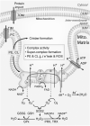
Role of mitochondrial phospholipids on mitochondrial function and ROS production. PE, PC, and CL exist in the IMM but in part due to their different shapes exert different effects on mitochondria form and function. For example, PE and CL are cone shaped and exert negative curvature on membranes which in the mitochondria serve to promote cristae formation. PC is cylindrical in shape, promoting lipid bilayer formation. In addition to the roles of these phospholipids to affect membrane properties, they have also been implicated in directly interacting with mitochondrial enzymes to influence their activities. For example, PC has been shown to interact with and augment the translocase of TIM suggesting that PC may be important for effective import of proteins into the mitochondrial matrix. Both PE and CL interact with and enhance the activities of several enzymes in the ETS. PE and CL also promote supercomplex formation that may facilitate efficient electron transfer. Electron leak occurs primarily at complex I and III and when combined with molecular oxygen forms the highly reactive ROS superoxide. To limit damage to the mitochondria, the mitochondrial matrix is equipped with SOD, which serves to convert superoxide to H2O2. H2O2, while less reactive than superoxide, is still damaging but is detoxified to water by several enzymes such as GPx, which utilizes reduced GSH as a co-factor, and the Trx/Prx antioxidant system, which utilizes NADPH as a co-factor. ETS, electron transport system; GPx, glutathione peroxidase; GSH, glutathione; H2O2, hydrogen peroxide; Prx, peroxiredoxin; ROS, reactive oxygen species; SOD, superoxide dismutase; TIM, the inner membrane; Trx, thioredoxin.
FIG. 3.
FIG. 3.
Unloading reduces mitochondrial phospholipid content, promotes mitochondrial ROS production and muscle atrophy. Muscle loading via regular physical activity and/or exercise promotes the abundance of PE and CL in muscle while attenuating the PC/PE ratio to promote mitochondrial function and decrease mitochondrial ROS production. These effects coincide with maintenance of muscle size and function. Conversely, muscle unloading via lack of physical activity or exercise reduces PE and CL content in muscle, impairing mitochondrial function, promoting mitochondrial ROS production and coincides with muscle atrophy and impaired muscle function.
FIG. 4.
FIG. 4.
Overview of lipid peroxidation propagation. Phospholipids containing PUFA are targeted by reactive oxygen species that undergo lipid radical propagation. This process begins with a hydroxyl radical reacting with a carbon double bond to form lipid radical molecules that have the capacity to react with a double bond of another phospholipid to propagate more lipid radicals. LOOH become degraded to form highly reactive carbonyl end products such as 4-HNE, MDA, and acrolein. LOOH, lipid peroxides; PUFA, polyunsaturated fatty acid.
FIG. 5.
FIG. 5.
Schematic overview of the LOOH pathway in skeletal muscle. Intracellular level of cysteine is tightly controlled by the system Xc (inhibited by erastin) at the plasma membrane. In turn, cysteine becomes metabolized to form GSH, a substrate for GPx4. GPx4 is the central enzyme in regulating the abundance of cellular LOOH, neutralizing them to nonreactive lipid alcohols. RSL3 is a commonly used inhibitor of GPx4. The Lands cycle plays an important role in incorporating phospholipids that contain polyunsaturated fatty acids (PUFA-PL). In particular, LPCAT3 has a high specificity to incorporate PUFA into a lyso-PL to form PUFA-PL. Lipoxygenases (ALOX) are responsible for the peroxidation of these PUFA-PLs. PLA2 works antagonistically to LPCAT3 and cleaves PUFAs, forming a lysophospholipid. Thus, inhibition of Lands cycle or LPCAT3 can be an effective strategy to suppress LOOH accumulation. COX increase LOOH through an alternative pathway converting AA to prostaglandins (PG). Ferrostatin-1 and liproxstatin-1 are inhibitors of LOOH accumulation with an inconclusive mechanism of action. We have utilized N-acetylcarnosine to successfully suppress muscle LOOH accumulation (Eshima et al, 2021). AA, arachidonic acid; COX, cyclooxygenases; LPCAT3, lysophosphatidylcholine acyltranserase 3; lyso-PL, lysophospholipid; PLA2, phospholipase A2.

Similar articles

Cited by

References

    1. Ahn B, Ranjit R, Premkumar P, et al. . Mitochondrial oxidative stress impairs contractile function but paradoxically increases muscle mass via fibre branching. J Cachexia Sarcopenia Muscle 2019;10(2):411–428; doi: 10.1002/jcsm.12375 - DOI - PMC - PubMed
    1. Aldini G, Orioli M, Rossoni G, et al. . The carbonyl scavenger carnosine ameliorates dyslipidaemia and renal function in Zucker obese rats. J Cell Mol Med 2011;15(6):1339–1354; doi: 10.1111/j.1582-4934.2010.01101.x - DOI - PMC - PubMed
    1. Anderson EJ, Vistoli G, Katunga LA, et al. . A carnosine analog mitigates metabolic disorders of obesity by reducing carbonyl stress. J Clin Invest 2018;128(12):5280–5293; doi: 10.1172/JCI94307 - DOI - PMC - PubMed
    1. Aranda R, Doménech E, Diana Rus A, et al. . Age-related increase in xanthine oxidase activity in human plasma and rat tissues. Free Radic Res 2007;41(11):1195–1200; doi: 10.1080/10715760701481461 - DOI - PubMed
    1. Arikketh D, Nelson R, Vance JE. Defining the importance of phosphatidylserine synthase-1 (PSS1). J Biol Chem 2008;283(19):12888–12897; doi: 10.1074/jbc.m800714200 - DOI - PubMed

Publication types

MeSH terms

LinkOut - more resources