. 2022 Nov 25;378(6622):eabo2523.
doi: 10.1126/science.abo2523. Epub 2022 Nov 25.

Unadjuvanted intranasal spike vaccine elicits protective mucosal immunity against sarbecoviruses

Affiliations

Unadjuvanted intranasal spike vaccine elicits protective mucosal immunity against sarbecoviruses

Tianyang Mao et al. Science. .

Abstract

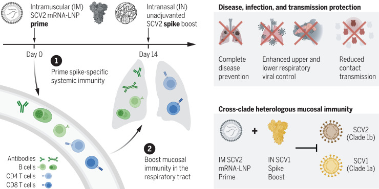
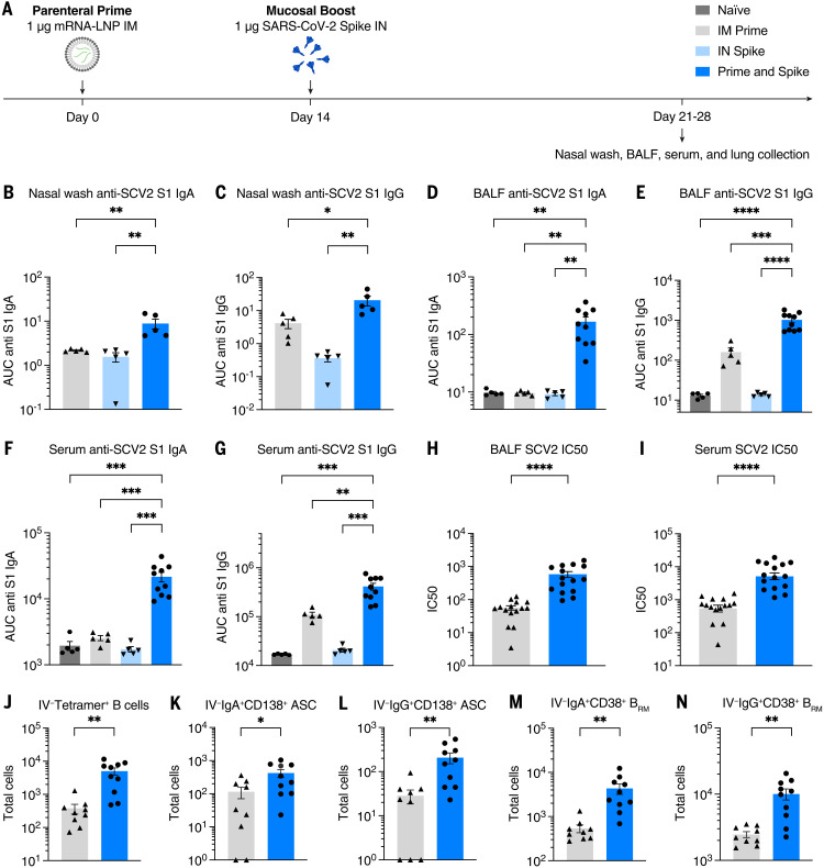
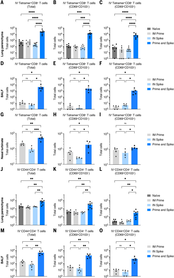
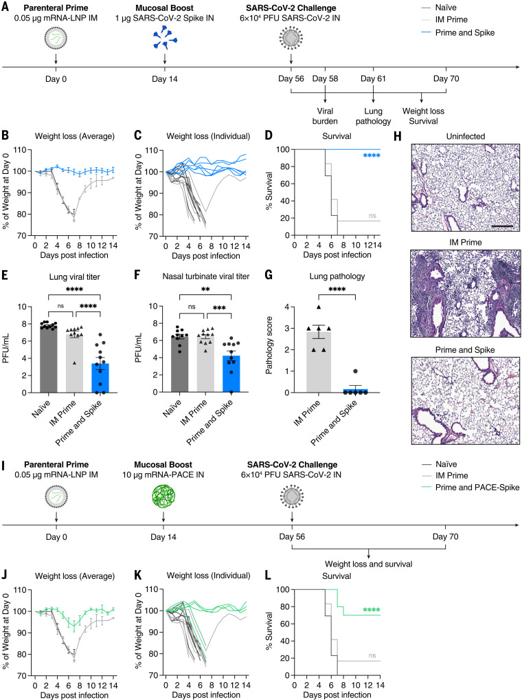
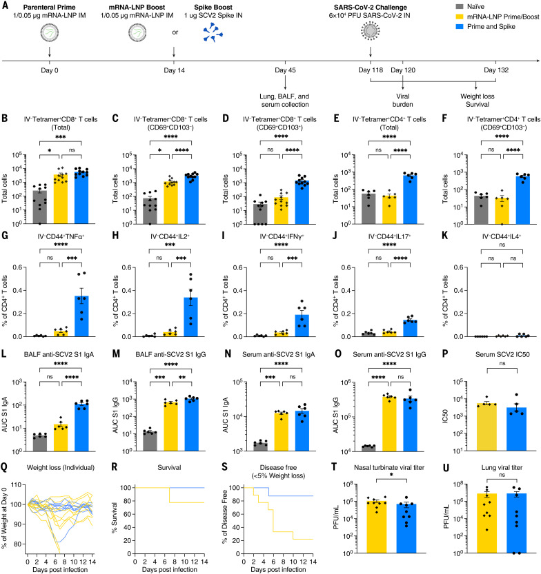
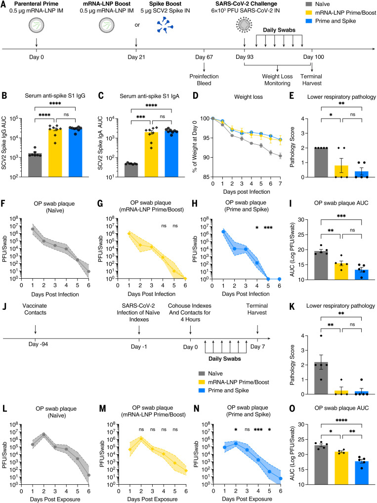
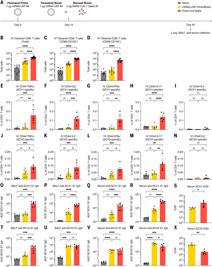
The severe acute respiratory syndrome coronavirus 2 (SARS-CoV-2) pandemic has highlighted the need for vaccines that not only prevent disease but also prevent transmission. Parenteral vaccines induce robust systemic immunity but poor immunity at the respiratory mucosa. We developed a vaccine strategy that we call "prime and spike," which leverages existing immunity generated by primary vaccination (prime) to elicit mucosal immune memory within the respiratory tract by using unadjuvanted intranasal spike boosters (spike). We show that prime and spike induces robust resident memory B and T cell responses, induces immunoglobulin A at the respiratory mucosa, boosts systemic immunity, and completely protects mice with partial immunity from lethal SARS-CoV-2 infection. Using divergent spike proteins, prime and spike enables the induction of cross-reactive immunity against sarbecoviruses.


Figures

None
A parenteral prime-unadjuvanted mucosal boost vaccine that elicits mucosal immunity against sarbecoviruses.
P&S converts systemic immunity generated by primary vaccination into local immunity in the respiratory mucosa. P&S affords protection against disease development, respiratory viral replication, and contact transmission after SARS-CoV-2 (SCV2) infection. Intranasal boosting by using a divergent spike protein from SARS-CoV-1 (SCV1) induces mucosal immunity against diverse sarbecovirus clades.
Fig. 1.
Fig. 1.. IN boosting with stabilized SARS-CoV-2 spike induces mucosal humoral memory.
(A) Experimental schema. Mice were intramuscularly immunized with 1 μg of mRNA-LNPs encoding full-length SARS-CoV-2 (SCV2) spike protein (Pfizer/BioNTech BNT162b2), followed by IN immunization with 1 μg of prefusion-stabilized (Hexapro), trimeric, recombinant SCV2 spike protein 14 days after mRNA-LNP immunization. Fourteen days after IN boost, serum, BALF, and nasal washes were collected to assess binding and neutralizing antibody responses. Lung tissues were collected for extravascular B cell analysis. (B to G) Measurement of SCV2 spike S1 subunit–specific (B) nasal wash IgA, (C) nasal wash IgG, (D) BALF IgA, (E) BALF IgG, (F) serum IgA, and (G) serum IgG in naïve mice, mice immunized with mRNA-LNP IM (IM Prime), mice immunized with the spike protein IN (IN Spike), or mice IM primed and IN boosted with spike (P&S). (H to K) Measurement of neutralization titer against SCV2 spike–pseudotyped vesicular stomatitis virus (VSV) in (H) BALF and (I) serum. (J to N) Using CD45 IV labeling, various extravascular (IV labeling antibody negative) B cell subsets were measured, including RBD tetramer-binding B cells, IgA+ BRM cells, IgG+ BRM cells, IgA+ ASCs, and IgG+ ASC in lung tissues from IM Prime or P&S mice. Mean ± SEM. Statistical significance was calculated by means of [(B) to (G)] one-way analysis of variance (ANOVA) or [(H) to (N)] Student’s t test; *P ≤ 0.05, **P ≤ 0.01, ***P ≤ 0.001, ****P ≤ 0.0001. Individual data points are represented and are pooled from two or three independent experiments.
Fig. 2.
Fig. 2.. IN boosting with stabilized SARS-CoV-2 spike induces mucosal T cell memory.
K18-hACE2 mice were intramuscularly primed with 1 μg mRNA-LNP and 14 days later intranasally boosted with 1 μg SCV2 spike. Lung tissues, BALF, and nasal turbinates were collected for extravascular T cell analysis. Lung tissues were collected 14 days after boost, whereas BALF and nasal turbinates were obtained 7 days after boost. (A to I) Extravascular CD8 T cell responses. Shown are quantification of SCV2 spike–specific tetramer+ CD8 T cells, CD69+CD103tetramer+ CD8 T cells, or CD69+CD103+tetramer+ CD8 T cells in [(A) to (C)] lung tissues, [(D) to (F)] BALF, or [(G) to (I)] nasal turbinates from naïve, IM prime, IN spike, or P&S mice. (J to O) Extravascular CD4 T cell responses. Shown are quantification of activated polyclonal CD4 T cells, CD69+CD103 CD4 T cells, or CD69+CD103+ CD4 T cells in [(J) to (L)] lung tissues or [(M) to (O)] BALF from naïve, IM prime, IN spike, or P&S mice. Mean ± SEM. Statistical significance was calculated by means of [(B) to (O)] one-way ANOVA followed by Tukey’s correction; *P ≤ 0.05, **P ≤ 0.01, ***P ≤ 0.001, ****P ≤ 0.0001. Individual data points are represented and are pooled from two or three independent experiments.
Fig. 3.
Fig. 3.. IN SARS-CoV-2 spike boosting protects against COVID-19–like disease.
(A) Experimental schema. K18-hACE2 mice were intramuscularly primed with 0.05 μg of mRNA-LNP and intranasally boosted with 1 μg of spike 14 days after IM prime. Six weeks after boost, mice were challenged with 6 × 10⁴ PFU SCV2 (2019n-CoV/USA_WA1/2020). The first cohort was used to evaluate weight loss and survival up to 14 days after infection. The second cohort was used to collect lung and nasal turbinate tissues 2 days after infection for viral titer measurement. The third cohort was used to collect lung tissues 5 days after infection for histological assessment. (B to D) Weight loss and survival of naïve, IM prime, or P&S mice from 1 to 14 days after infection. (E to F) Measurement of infectious virus titer in lung and nasal turbinate tissues at 2 days after infection by means of plaque assay. (G) Pathology score of lung sections at 5 days after infection by means of H&E staining. (H) Representative H&E staining results from uninfected, IM prime, or P&S mice. Scale bar, 250 μm. Sections are representative of multiple sections from at least five mice per group. (I) Experimental schema. K18-hACE2 mice were intramuscularly primed with 0.05 μg of mRNA-LNP and intranasally boosted with 10 μg of mRNA encapsulated by PACE (IN PACE-Spike) 14 days after IM Prime. Six weeks after boost, mice were challenged with 6 × 10⁴ PFU SCV2 (2019n-CoV/USA_WA1/2020). Weight loss and survival were monitored up to 14 days after infection. (J to L) Weight loss and survival of naïve, IM prime, or prime and PACE-spike K18-hACE2 mice from 1 to 14 days after infection. Mean ± SEM. Statistical significance was calculated by means of [(D) and (L)] log-rank Mantel-Cox test, [(E) and (F)] one-way ANOVA followed by Tukey’s correction, or (G) Student’s t test; *P ≤ 0.05, **P ≤ 0.01, ***P ≤ 0.001, ****P ≤ 0.0001. Individual data points are represented and are pooled from two independent experiments.
Fig. 4.
Fig. 4.. IN spike boosting elicits enhanced mucosal immunity with similar systemic humoral responses to IM mRNA-LNP boosting.
(A) Experimental schema. K18-hACE2 mice were IM primed with 1 μg of mRNA-LNP, followed 14 days later by boosting with 1 μg of mRNA-LNP IM or 1 μg of SCV2 spike IN. Forty-five days after prime, lung tissues were collected for T cell analysis by means of flow cytometry, and BALF and serum were collected for antibody measurement. K18-hACE2 mice were intramuscularly primed with 0.05 μg of mRNA-LNP, followed 14 days later by boosting with 0.05 μg of mRNA-LNP intramuscularly, or 1 μg of SCV2 spike intranasally and challenged with 6 × 10⁴ PFU SCV2 at 118 days after prime. (B to D) Quantification of total tetramer+ CD8 T cells, CD69+CD103tetramer+ CD8 T cells, or CD69+CD103+tetramer+ CD8 T cells in lung tissues from naïve, mRNA-LNP prime-boost, or P&S mice. (E to F) Quantification of total tetramer+ CD4 T cells or CD69+CD103tetramer+ CD4 T cells in lung tissues. (G to K) Lung lymphocytes were isolated by means of Percoll gradient and restimulated with spike peptide megapool from SCV2. Intracellular cytokine staining was performed to assess antigen-specific production of TNF-α, IL-2, IFN-γ, IL-17, and IL-4 by extravascular IV-CD45CD44+ CD4 T cells. (L to O) Measurement of SCV2 spike S1 subunit–specific (L) BALF IgA, (M) BALF IgG, (N) serum IgA, and (O) serum IgG in naïve, mRNA-LNP prime-boost, or P&S mice. (P) Measurement of neutralization titer against SCV2 spike–pseudotyped VSV. (Q to S) Weight loss, survival, and disease-free survival (<5% maximum weight loss) of mRNA-LNP prime-boost or P&S mice from 1 to 14 days after infection. (T and U) Measurement of infectious virus titer in lung and nasal turbinate tissues at 2 days after infection by means of plaque assay. To reduce overall number of experimental animals used, control data points from naïve and mRNA prime-boost are common to Figs. 4 and 6. Mean ± SEM. Statistical significance was calculated by means of [(B) to (O)] one-way ANOVA followed by Tukey’s correction or [(P), (T), and (U)] Student’s t test, and [(R) and (S)] log-rank Mantel-Cox test; *P ≤ 0.05, **P ≤ 0.01, ***P ≤ 0.001, ****P ≤ 0.0001. Individual data points are represented and are pooled from two independent experiments.
Fig. 5.
Fig. 5.. IN spike boosting leads to reduced viral transmission in hamster model.
(A) Experimental schema. Syrian hamsters were intramuscularly primed with 0.5 μg of mRNA-LNP, followed 21 days later by boosting with 0.5 μg of mRNA-LNP intramuscularly or 5 μg of SCV2 spike intranasally. (B and C) Sixty-seven days after prime, serum IgG and IgA were assessed by means of ELISA. At 93 days after prime, naïve, mRNA-LNP prime-boost, and P&S hamsters were infected with 6 × 10³ PFU SCV2. (D) Weight loss as percent of starting. (E) Histopathologic analysis of lung samples at 7 days after infection. (F to H) Viral titer from oropharyngeal swabs are shown as mean (symbols) and standard deviation (shaded regions), P value relative to control at the same time point. (I) AUC analysis for viral titer over 6 days after infection. (J) Transmission experimental schema. Syrian hamsters vaccinated as above were cohoused for 4 hours with naïve donor hamsters that had been infected 24 hours earlier with 6 × 10³ PFU SCV2. (K) Histopathologic analysis of lung samples at 7 days after exposure. (L to N) Viral titers from oropharyngeal swabs are shown as mean (symbols) and standard deviation (shade), P value relative to control at the same time point. (O) AUC analysis for viral titer over 6 days after infection. Mean ± SEM. Statistical significance was calculated by means of [(B), (C), (E), (I), (K), and (O)] one-way ANOVA followed by Tukey’s correction, [(F) to (H)] mixed-effect analysis followed by Tukey’s multiple comparison test, or [(L) to (N)] two-way ANOVA followed by Dunnett’s multiple comparisons test; *P ≤ 0.05, **P ≤ 0.01, ***P ≤ 0.001, ****P ≤ 0.0001. Individual data points are represented from one independent experiment.
Fig. 6.
Fig. 6.. Heterologous IN boosting with SARS-CoV-1 spike enhances preexisting SCV2-specific immunity and broadens reactivities to SCV1.
(A) Experimental schema. K18-hACE2 mice were intramuscularly primed with 1 μg of mRNA-LNP, followed by boosting with 1 μg of mRNA-LNP intramuscularly, or 5 μg of prefusion-stabilized, trimeric, recombinant SARS-CoV-1 (SCV1) spike IN (IN SpikeX) 14 days after prime. (B to D) Quantification of total tetramer+ CD8 T cells, CD69+CD103tetramer+ CD8 T cells, or CD69+CD103+tetramer+ CD8 T cells in lung tissues from naïve, mRNA-LNP prime-boost, or P&Sx mice. (E to N) Percoll gradient purified lung lymphocytes were restimulated with spike peptide megapool from [(E) to (I)] SCV1 or [(J) to (N)] SCV2, and intracellular cytokine staining was performed to assess antigen-specific production of TNF-α, IL-2, IFN-γ, IL-17, and IL-4 by extravascular IV-CD45CD44+ CD4 T cells. (O to S) Measurement of SCV1 spike S1 subunit–specific BALF IgA and IgG, and serum IgA and IgG. (S) Measurement of neutralization titer against SCV1 spike–pseudotyped VSV. (T to W) Measurement of SCV2 spike S1 subunit–specific BALF IgA and IgG, and serum IgA and IgG. (X) Measurement of neutralization titer against SCV2 spike–pseudotyped VSV. To reduce overall number of experimental animals used, control data points from naïve and mRNA prime-boost are common to Figs. 4 and 6. Mean ± SEM. Statistical significance was calculated by means of one-way ANOVA followed by Tukey’s correction, except for [(S) and (X)] Student’s t test; *P ≤ 0.05, **P ≤ 0.01, ***P ≤ 0.001, ****P ≤ 0.0001. Individual data points are represented and are pooled from two independent experiments.

Update of

References

    1. Baden L. R., El Sahly H. M., Essink B., Kotloff K., Frey S., Novak R., Diemert D., Spector S. A., Rouphael N., Creech C. B., McGettigan J., Khetan S., Segall N., Solis J., Brosz A., Fierro C., Schwartz H., Neuzil K., Corey L., Gilbert P., Janes H., Follmann D., Marovich M., Mascola J., Polakowski L., Ledgerwood J., Graham B. S., Bennett H., Pajon R., Knightly C., Leav B., Deng W., Zhou H., Han S., Ivarsson M., Miller J., Zaks T., COVE Study Group , Efficacy and safety of the mRNA-1273 SARS-CoV-2 vaccine. N. Engl. J. Med. 384, 403–416 (2021). 10.1056/NEJMoa2035389 - DOI - PMC - PubMed
    1. Dagan N., Barda N., Kepten E., Miron O., Perchik S., Katz M. A., Hernán M. A., Lipsitch M., Reis B., Balicer R. D., BNT162b2 mRNA COVID-19 vaccine in a nationwide mass vaccination setting. N. Engl. J. Med. 384, 1412–1423 (2021). 10.1056/NEJMoa2101765 - DOI - PMC - PubMed
    1. Emary K. R. W., Golubchik T., Aley P. K., Ariani C. V., Angus B., Bibi S., Blane B., Bonsall D., Cicconi P., Charlton S., Clutterbuck E. A., Collins A. M., Cox T., Darton T. C., Dold C., Douglas A. D., Duncan C. J. A., Ewer K. J., Flaxman A. L., Faust S. N., Ferreira D. M., Feng S., Finn A., Folegatti P. M., Fuskova M., Galiza E., Goodman A. L., Green C. M., Green C. A., Greenland M., Hallis B., Heath P. T., Hay J., Hill H. C., Jenkin D., Kerridge S., Lazarus R., Libri V., Lillie P. J., Ludden C., Marchevsky N. G., Minassian A. M., McGregor A. C., Mujadidi Y. F., Phillips D. J., Plested E., Pollock K. M., Robinson H., Smith A., Song R., Snape M. D., Sutherland R. K., Thomson E. C., Toshner M., Turner D. P. J., Vekemans J., Villafana T. L., Williams C. J., Hill A. V. S., Lambe T., Gilbert S. C., Voysey M., Ramasamy M. N., Pollard A. J., COVID-19 Genomics UK consortium, AMPHEUS Project, Oxford COVID-19 Vaccine Trial Group , Efficacy of ChAdOx1 nCoV-19 (AZD1222) vaccine against SARS-CoV-2 variant of concern 202012/01 (B.1.1.7): An exploratory analysis of a randomised controlled trial. Lancet 397, 1351–1362 (2021). 10.1016/S0140-6736(21)00628-0 - DOI - PMC - PubMed
    1. Shah A. S. V., Gribben C., Bishop J., Hanlon P., Caldwell D., Wood R., Reid M., McMenamin J., Goldberg D., Stockton D., Hutchinson S., Robertson C., McKeigue P. M., Colhoun H. M., McAllister D. A., Effect of vaccination on transmission of SARS-CoV-2. N. Engl. J. Med. 385, 1718–1720 (2021). 10.1056/NEJMc2106757 - DOI - PMC - PubMed
    1. Harris R. J., Hall J. A., Zaidi A., Andrews N. J., Dunbar J. K., Dabrera G., Effect of vaccination on household transmission of SARS-CoV-2 in England. N. Engl. J. Med. 385, 759–760 (2021). 10.1056/NEJMc2107717 - DOI - PMC - PubMed

Publication types