

doi: 10.1016/j.clinimag.2022.10.008.
Epub 2022 Oct 20.
Imaging features of anorectal proctitis in monkeypox infection
Affiliations
- PMID: 36302321
- PMCID: PMC9583688
- DOI: 10.1016/j.clinimag.2022.10.008
Item in Clipboard
Imaging features of anorectal proctitis in monkeypox infection
Clin Imaging.
2022 Dec.
Abstract
The monkeypox outbreak of 2022 saw the first community-sustained transmission of the monkeypox virus outside of Africa, and rapidly developed into multi-country spread. A common presenting sign of monkeypox infection during this outbreak has been rectal pain due to proctitis. Proctitis with large hypoattenuated anorectal ulcers on CT scan should invoke consideration for monkeypox infection in young homosexual or bisexual men with associated skin eruptions.
Keywords: Anorectal ulceration; Monkeypox; Proctitis.
Copyright © 2022 Elsevier Inc. All rights reserved.
Conflict of interest statement
Declaration of competing interest The authors have no conflicts of interest to declare.
Figures
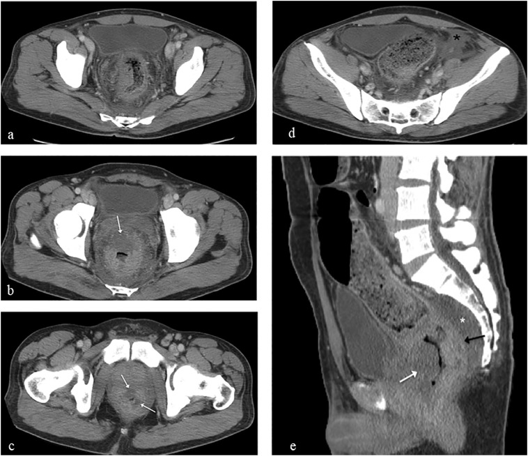
Axial images from contrast-enhanced CT scan of the abdomen and pelvis reveal (a) diffuse circumferential rectal mural thickening, hazy infiltrated perirectal fat, and presacral edema with discrete geographic non-enhancing hypoattenuated zones (white arrows) in the rectal (b) and anal wall (c) due to large ulcerations and (d) pelvic ascites (black asterisk). Several bilateral inguinal lymph nodes measuring up to 1 cm are seen on (c). Sagittal image (e) reveals the thickened enhancing posterior rectal wall (black arrow) contrasting a large anterior wall hypoattenuated ulcer (white arrow). Note prominent presacral edema (white asterisk). A small right pleural effusion is not shown.
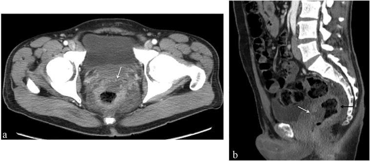
Axial image from contrast-enhanced CT scan 11 days later (a) shows decrease in mural thickening and anterior hypoattenuated ulcer. Sagittal image (b) shows decrease in enhancing posterior rectal wall (black arrow) and large anterior wall hypoattenuated ulcer (white arrow) compared with prior exam. Pelvic ascites resolved (not shown).
Similar articles
-
CT findings of proctitis in two patients with Mpox.Emerg Radiol. 2023 Jun;30(3):387-389. doi: 10.1007/s10140-023-02140-x. Epub 2023 May 10. Emerg Radiol. 2023. PMID: 37162597
-
Monkeypox outbreak in Spain: clinical and epidemiological findings in a prospective cross-sectional study of 185 cases.Br J Dermatol. 2022 Nov;187(5):765-772. doi: 10.1111/bjd.21790. Epub 2022 Aug 20. Br J Dermatol. 2022. PMID: 35917191
-
Overlooked monkeypox cases among men having sex with men during the 2022 outbreak - a retrospective study.Int J Infect Dis. 2023 Mar;128:58-60. doi: 10.1016/j.ijid.2022.12.014. Epub 2022 Dec 16. Int J Infect Dis. 2023. PMID: 36529372 Free PMC article.
-
Monkeypox: an epidemiologic and clinical comparison of African and US disease.J Am Acad Dermatol. 2006 Sep;55(3):478-81. doi: 10.1016/j.jaad.2006.05.061. J Am Acad Dermatol. 2006. PMID: 16908354 Free PMC article. Review.
-
The Historical Epidemiology of Human Monkeypox: A Review of Evidence from the 1970 Emergence to the 2022 Outbreak.Tohoku J Exp Med. 2022 Nov 19;258(4):243-255. doi: 10.1620/tjem.2022.J081. Epub 2022 Oct 6. Tohoku J Exp Med. 2022. PMID: 36198504 Review.
Cited by
-
CT findings of proctitis in two patients with Mpox.Emerg Radiol. 2023 Jun;30(3):387-389. doi: 10.1007/s10140-023-02140-x. Epub 2023 May 10. Emerg Radiol. 2023. PMID: 37162597
-
Navigating monkeypox: identifying risks and implementing solutions.Open Vet J. 2024 Dec;14(12):3144-3163. doi: 10.5455/OVJ.2024.v14.i12.1. Epub 2024 Dec 31. Open Vet J. 2024. PMID: 39927376 Free PMC article. Review.
-
Proctitis in patients with monkeypox infection: a single-center analysis of 42 consecutive cases from a multidisciplinary observational study on monkeypox proctitis.Tech Coloproctol. 2023 Dec;27(12):1211-1218. doi: 10.1007/s10151-023-02782-6. Epub 2023 Apr 22. Tech Coloproctol. 2023. PMID: 37086291 Free PMC article.
-
Mpox gastrointestinal manifestations: a systematic review.BMJ Open Gastroenterol. 2024 Jan 6;11(1):e001266. doi: 10.1136/bmjgast-2023-001266. BMJ Open Gastroenterol. 2024. PMID: 38184298 Free PMC article.
-
Monkeypox in a patient with HIV: case report.Rev Peru Med Exp Salud Publica. 2023 Apr-Jun;40(2):229-235. doi: 10.17843/rpmesp.2023.402.12344. Rev Peru Med Exp Salud Publica. 2023. PMID: 38232270 Free PMC article.
References
-
- Center for Disease Control and Prevention, National Center for Emerging and Zoonotic Infectious Diseases, Division of High-Consequence Pathogens and Pathology. 2022 Monkeypox Outbreak Global Map. United States Department of Health and Human Services. Accessed 20 September 2022, https://www.cdc.gov/poxvirus/monkeypox/response/2022/world-map.html.
-
- World Health Organization Surveillance, case investigation and contact tracing for monkeypox: interim guidance. WHO Health Emergencies Programme. 25 August 2022:1–12. https://www.who.int/publications/i/item/WHO-MPX-Surveillance-2022.3
MeSH terms
LinkOut - more resources
Full Text Sources
Medical
