Sagittal Plane Geometry of Cervical, Thoracic, and Lumbar Endplates
- PMID: 36302610
- PMCID: PMC10168012
- DOI: 10.14444/8336
Sagittal Plane Geometry of Cervical, Thoracic, and Lumbar Endplates
Abstract
Background: Many studies have emphasized the importance of interface contact between implants and the vertebral endplate (VE). The goal of this study was to analyze the shape and other specific parameters of the VE to provide reference data for better implant interface contact in intervertebral disc space procedures.
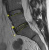
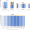
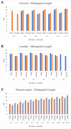
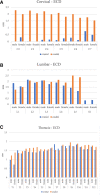
Methods: Cervical, thoracic, and lumbar spine midsagittal plane magnetic resonance images of 100 adults (58 women) were analyzed. The morphology of the VEs was classified as concave, convex, flat, or irregular. Midsagittal endplate length (ML), endplate concavity depth (ECD), and endplate concavity axis (ECA) location were measured in the midsagittal plane. The parameters were compared between the cervical, thoracic, and lumbar spines and between the sexes.
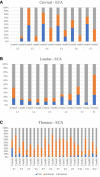
Results: The VE morphology, ML, ECD, and ECA showed variations along the spine, mainly in the cervical and lower lumbar spines. The sagittal geometry of the VE was not flat or uniform along the cervical, thoracic, and lumbar spines. Different morphological types were observed along different spinal segments and according to sex. In the cervical spine, the majority of cranial VEs were flat, while caudal VEs were mostly concave.
Conclusion: Sagittal VE geometry should be taken into consideration during the use of intervertebral cages or disc arthroplasty.
Keywords: MRI; cervical; endplate; lumbar spine; sagittal geometry; thoracic.
This manuscript is generously published free of charge by ISASS, the International Society for the Advancement of Spine Surgery. Copyright © 2022 ISASS. To see more or order reprints or permissions, see http://ijssurgery.com.
Conflict of interest statement
Declaration of Conflicting Interests: The authors report no conflicts of interest in this work.
Figures

Similar articles
-
Geometry of inferior endplates of the cervical spine.Clin Neurol Neurosurg. 2016 Mar;142:132-136. doi: 10.1016/j.clineuro.2016.01.027. Epub 2016 Jan 25. Clin Neurol Neurosurg. 2016. PMID: 26852320
-
Sagittal endplate morphology of the lower lumbar spine.Eur Spine J. 2012 May;21 Suppl 2(Suppl 2):S160-4. doi: 10.1007/s00586-012-2168-4. Eur Spine J. 2012. PMID: 22315035 Free PMC article.
-
Association Between Measures of Vertebral Endplate Morphology and Lumbar Intervertebral Disc Degeneration.Can Assoc Radiol J. 2017 May;68(2):210-216. doi: 10.1016/j.carj.2016.11.002. Epub 2017 Feb 16. Can Assoc Radiol J. 2017. PMID: 28216287
-
Effects of sagittal endplate shape on lumbar segmental mobility as evaluated by kinetic magnetic resonance imaging.Spine (Phila Pa 1976). 2014 Aug 1;39(17):E1035-41. doi: 10.1097/BRS.0000000000000419. Spine (Phila Pa 1976). 2014. PMID: 24859573
-
Sagittal geometry of the middle and lower cervical endplates.Eur Spine J. 2013 Jul;22(7):1570-5. doi: 10.1007/s00586-013-2791-8. Epub 2013 Apr 24. Eur Spine J. 2013. PMID: 23612902 Free PMC article.
Cited by
-
The footprint mismatch of cervical disc arthroplasty comes from degenerative factor besides ethnic factor.Sci Rep. 2024 Sep 5;14(1):20673. doi: 10.1038/s41598-024-71786-5. Sci Rep. 2024. PMID: 39237767 Free PMC article.
-
Spinal Disease Burden and Priorities of Community Spine Care in the Brazilian Public Health System vs the United States.Int J Spine Surg. 2023 Jun;17(3):477-481. doi: 10.14444/8452. Epub 2023 Jun 2. Int J Spine Surg. 2023. PMID: 37268431 Free PMC article. No abstract available.
References
LinkOut - more resources
Full Text Sources
