Review
. 2022 Oct 27;8(1):69.
doi: 10.1038/s41572-022-00398-y.

Gallbladder cancer

Affiliations
Review

Gallbladder cancer

Juan C Roa et al. Nat Rev Dis Primers. .

Erratum in

  • Publisher Correction: Gallbladder cancer.
    Roa JC, García P, Kapoor VK, Maithel SK, Javle M, Koshiol J. Roa JC, et al. Nat Rev Dis Primers. 2022 Nov 18;8(1):75. doi: 10.1038/s41572-022-00408-z. Nat Rev Dis Primers. 2022. PMID: 36400795 No abstract available.

Abstract

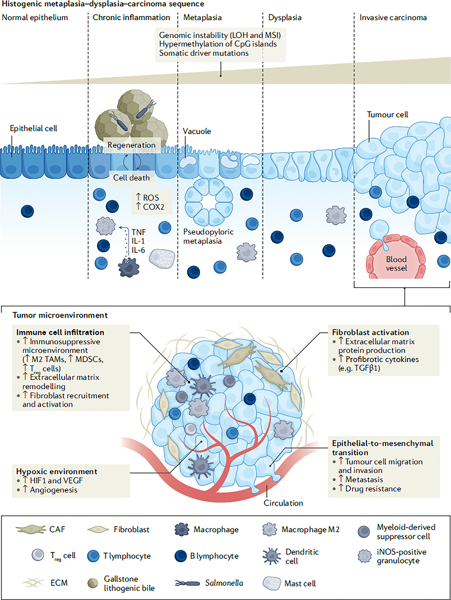
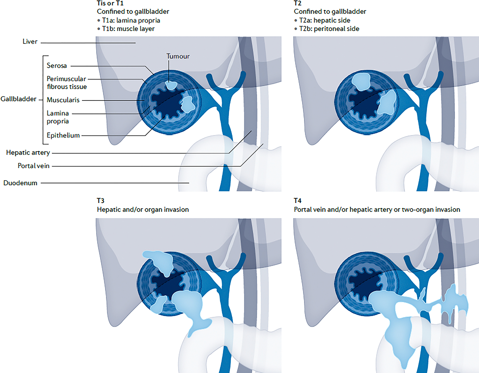
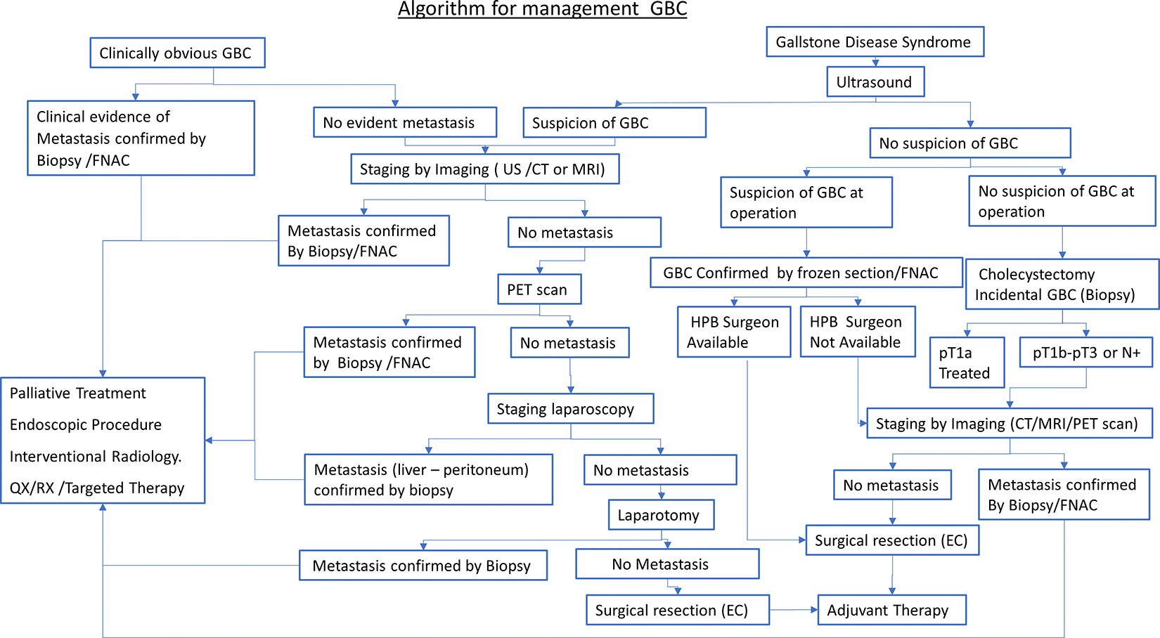
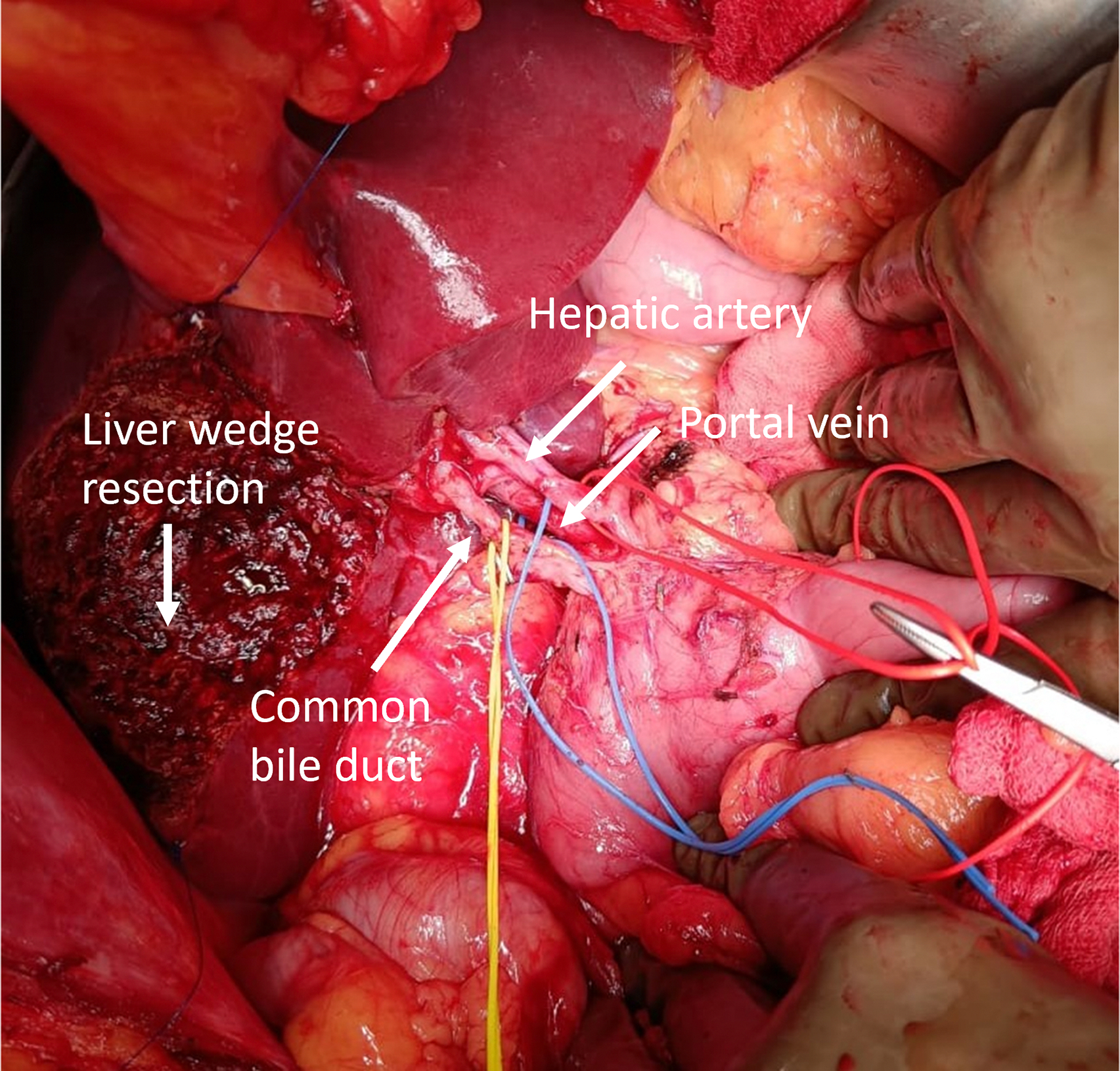
Gallbladder cancer (GBC) is the most common cancer of the biliary tract, characterized by a very poor prognosis when diagnosed at advanced stages owing to its aggressive behaviour and limited therapeutic options. Early detection at a curable stage remains challenging because patients rarely exhibit symptoms; indeed, most GBCs are discovered incidentally following cholecystectomy for symptomatic gallbladder stones. Long-standing chronic inflammation is an important driver of GBC, regardless of the lithiasic or non-lithiasic origin. Advances in omics technologies have provided a deeper understanding of GBC pathogenesis, uncovering mechanisms associated with inflammation-driven tumour initiation and progression. Surgical resection is the only treatment with curative intent for GBC but very few cases are suitable for resection and most adjuvant therapy has a very low response rate. Several unmet clinical needs require to be addressed to improve GBC management, including discovery and validation of reliable biomarkers for screening, therapy selection and prognosis. Standardization of preneoplastic and neoplastic lesion nomenclature, as well as surgical specimen processing and sampling, now provides reproducible and comparable research data that provide a basis for identifying and implementing early detection strategies and improving drug discovery. Advances in the understanding of next-generation sequencing, multidisciplinary care for GBC, neoadjuvant and adjuvant strategies, and novel systemic therapies including chemotherapy and immunotherapies are gradually changing the treatment paradigm and prognosis of this recalcitrant cancer.


Conflict of interest statement

Competing interests

M.J. receives research funding (to Institution) from Merck, EMD Serono, Novartis, Eli Lilly, Astra Zeneca, Genentech, Transthera, Meclun, BMS, Incyte, QED, Taiho, Servier, Oncosil, Basilea, Nucana; and to self or as advisory board/Data and safety Monitoring Board member from Incyte, Zymeworks, Mundi Pharma, Nucana, MORE health and Origimed. Peer grant funding from Department of Defenseand National Institute of Health. M.S. receives research funding (to Institution) from BMS, QED/HELSINN, Natera. The other authors (J.C.R., P.G., J.K., V.K.) declare no competing interests.

Figures

Figure 1.
Figure 1.. Gallbladder anatomy and histology
The gallbladder is located adhered under the right lobe of the liver mostly revested by visceral peritoneum, connected to the main biliary duct in the hepatic pedicle through the cystic duct. Gallbladder histology highlights the absence of muscularis mucosa and the discontinuity of the muscle layer, which are relevant to understanding difficulties in pathologic tumour staging.
Figure 2.
Figure 2.. Worldwide incidence of GBC
Estimated global age-standardized incidence rates (ASR) per 100,000 individuals of gallbladder cancer in 2020, in both sexes and all ages. Adapted from the International Agency for Research on Cancer’s GLOBOCAN database.
Figure 3.
Figure 3.. Model of gallbladder cancer progression
Most GBCs develop in the setting of cholelithiasis following a metaplasia-dysplasia-carcinoma sequence. Gallstones and proinflammatory lithogenic bile, and possibly bacterial infections, enable continuous cycles of epithelium destruction and regeneration leading to a long-standing chronic inflammatory response that promotes malignant transformation and tumour progression. Epithelial cells are shown as different colours to represent the non-homogenous transitions during carcinogenesis. Chronic cholecystitis is characterized by overexpression of cyclooxygenase-2 (COX-2) and high infiltration levels of T cells, B cells, macrophages expressing COX-2 and inducible nitric oxide synthase (iNOS), iNOS-positive granulocytes, and mast cells. Morphological changes are accompanied by genetic and epigenetic events that generally accumulate and persist at advanced stages. Genomic instability and TP53 alterations are recognized as the earliest genetic events, but loss-of-function of other tumour suppressor genes, such as CDKN2A, CDH1 and APC, has also been reported in chronic cholecystitis. Epigenetic alterations, predominantly hypermethylation of CpG island and promoter regions, also increase during GBC carcinogenesis. Once the tumour is established, several key cancer-related signaling pathways are dysregulated by genetic and/or epigenetic mechanisms, including the ErbB, Ras–MAPK, PI3K–AKT–mTOR, and Notch pathways. All these molecular alterations, along with the crosstalk between tumour and stromal cells, contribute to tumour progression by driving invasion and metastasis. More-aggressive GBCs are characterized by a mesenchymal-like phenotype and an immunosuppressive tumour microenvironment, showing upregulation of gene sets related to epithelial-to-mesenchymal transition, TGF-β signaling and immune pathways (B cell receptor signaling and cytokine-cytokine receptor interaction). Major players in the gallbladder tumour microenvironment, increased in poor-survival GBCs, include cancer associated fibroblasts (CAFs), endothelial cells and diverse immune cell populations, such as myeloid-derived suppressor cells (MDSCs) and M2-like tumour-associated macrophages (TAMs), which exert potent immunosuppressive activity. M2 TAMs: M2 tumour-associated macrophages; MSI: microsatellite instability; LOH: loss of heterozygosity; ECM: extracellular matrix; ROS: reactive oxygen species.
Figure 4.
Figure 4.. Metaplasia–dysplasia–carcinoma sequence
This histogenic sequence is frequently linked to gallstone inflammation elicited over a period of 15–25 years starting with multiple cycles of epithelial and mucosa necrosis regeneration, with atrophy and consequential intestinal, foveolar or pseudopyloric metaplasia. Low-grade and high-grade dysplasia follows the metaplasia stage and precedes intramucosal and muscle invasive carcinoma (early carcinoma), which are usually detected incidentally. Subserosal pT2 and serosal or hepatic pT3-T4 carcinomas are advanced stage disease and have poorer prognosis than pT1–2. Haematoxylin and eosin staining; magnification 10–20X.
Figure 5.
Figure 5.. GBC T-stage
The pathologic tumour (pT) stage of GBC is one of the most important prognostic factors, but it is also critical for treatment decision-making. Early cancers (pT1a and pT1b) are limited to the mucosa and muscle layer. Advanced cancers (pT2, pT2 and pT4) involve periconnective tissue of the gallbladder (pT2), gallbladder serosa, liver and/or one adjacent digestive structure such as stomach or duodenum (pT3), or infiltration of the hepatic pedicle, or two or more structures (pT4).
Figure 6.
Figure 6.. Adenoma–carcinoma sequence
This histogenic pathway is a less frequent sequence and probably associated with inflammation that is not related to cholelithiasis, such as in the setting of and anomalous pancreaticobiliary junction. WHO has recently renamed and classified these lesions into new entities (intracholecystic papillary-tubular neoplasms; ICNs) according to the predominant morphological features and cell linages (biliary, intestinal and tubular non-mucinous patterns) and their biological behavior according to different rates of association with invasive carcinoma. Haematoxylin and eosin staining; magnification 10–20X.
Figure 7.
Figure 7.. Algorithm for management of GBC
GBC usually presents as a flat lesion associated with cholelithiasis. Preoperative diagnosis by imaging is infrequent, as GBC cases are predominantly found during histopathologic examination (incidental GBC; blue boxes). When a localized GBC is suspected during surgery and a HPB surgeon is not available for an extended cholecystectomy a post-surgical staging by imaging is mandatory. When GBC is suspected preoperatively, metastatic disease has to be excluded by imaging before surgery. If metastatic disease is confirmed by biopsy and/or cytology, palliative management is required. FNA: Fine needle aspiration citology; HPB: Hepato pancreatico biliary; QX: Quemotherapy; Rx: Radiotherapy.
Figure 8.
Figure 8.. Extended cholecystectomy
Operative image shows the cut surface of the liver after non-anatomical liver wedge resection. Lymphadenectomy has been performed in the hepatoduodenal ligament baring the common bile duct (yellow sling), portal vein (blue sling) and the hepatic artery (red sling). (Image courtesy of Vinay Mahala, HPB Surgery MGMCH, Jaipur Rajasthan India).

Similar articles

Cited by

References

    1. Roa I, Ibacache G, Munoz S & de Aretxabala X Gallbladder cancer in Chile: Pathologic characteristics of survival and prognostic factors: analysis of 1,366 cases. Am J Clin Pathol 141, 675–682, doi: 10.1309/AJCPQT3ELN2BBCKA (2014). - DOI - PubMed
    1. Kapoor VK Gallbladder neck cancer and perihilar cholangiocarcinoma - siblings, cousins or look alikes? Korean J Hepatobiliary Pancreat Surg 19, 86–88, doi: 10.14701/kjhbps.2015.19.2.86 (2015). - DOI - PMC - PubMed
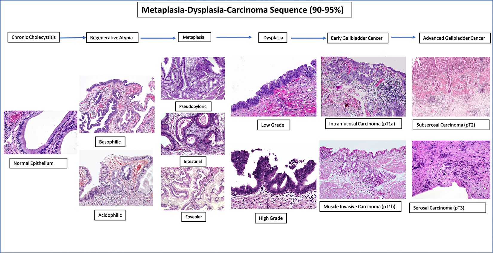
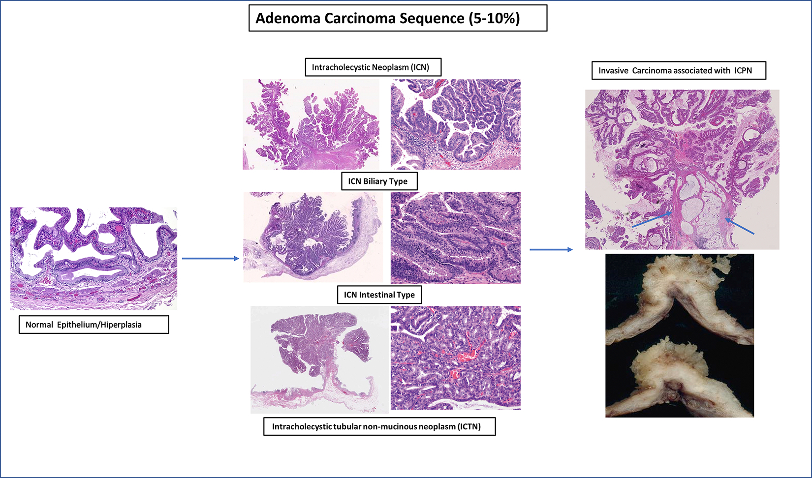
    1. Roa JC, Basturk O & Adsay V Dysplasia and carcinoma of the gallbladder: pathological evaluation, sampling, differential diagnosis and clinical implications. Histopathology 79, 2–19, doi: 10.1111/his.14360 (2021).

      Complete review that sets the basis for morphology nomenclature of preneoplastic and neoplastic gallbladder lesions.

    1. Roa J, Adsay NV, Arola J, Tsu W & Zen Y in Digestive System Tumours Vol. 1 WHO Classification of Tumours. (ed WHO Classification of Tumours Editorial Board; ) Ch. 9, 283–288 (IARC, 2019).
    1. Sharma A, Sharma KL, Gupta A, Yadav A & Kumar A Gallbladder cancer epidemiology, pathogenesis and molecular genetics: Recent update. World journal of gastroenterology 23, 3978–3998, doi: 10.3748/wjg.v23.i22.3978 (2017). - DOI - PMC - PubMed

Publication types

LinkOut - more resources