. 2022 Oct 27;5(1):1138.
doi: 10.1038/s42003-022-04068-3.

SARS-CoV-2 Omicron variant is attenuated for replication in a polarized human lung epithelial cell model

Affiliations

SARS-CoV-2 Omicron variant is attenuated for replication in a polarized human lung epithelial cell model

Christin Mache et al. Commun Biol. .

Abstract

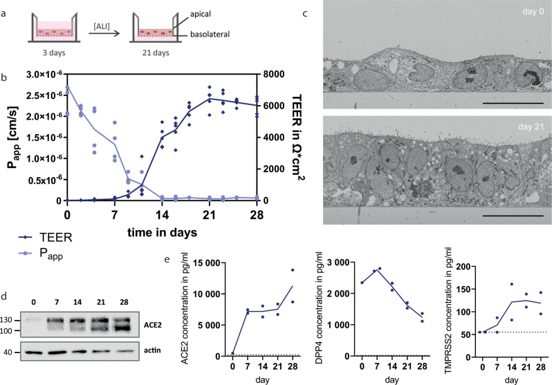
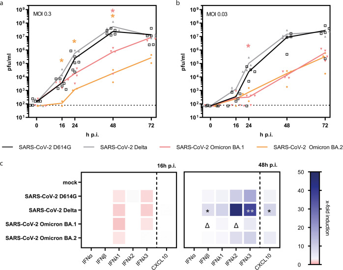
SARS-CoV-2 and its emerging variants of concern remain a major threat for global health. Here we introduce an infection model based upon polarized human Alveolar Epithelial Lentivirus immortalized (hAELVi) cells grown at the air-liquid interface to estimate replication and epidemic potential of respiratory viruses in the human lower respiratory tract. hAELVI cultures are highly permissive for different human coronaviruses and seasonal influenza A virus and upregulate various mediators following virus infection. Our analysis revealed a significantly reduced capacity of SARS-CoV-2 Omicron BA.1 and BA.2 variants to propagate in this human model compared to earlier D614G and Delta variants, which extends early risk assessments from epidemiological and animal studies suggesting a reduced pathogenicity of Omicron.


Conflict of interest statement

The authors declare no competing interests.

Figures

Fig. 1
Fig. 1. hAELVi cell air–liquid-interface cultures as model of the distal lung.
a Schematic representation of hAELVi air–liquid-interface (ALI) cultures. b Measurement of transepithelial electrical resistance (TEER) and permeability of sodium fluorescein across the cellular monolayer of hAELVi cells growing under ALI up to 28 days. Experiments were performed for five (TEER) or four (Papp) independent experiments in technical duplicates, respectively. c Electron microscopic analysis of hAELVi cells grown at the ALI for 0 or 21 days. Data are representative for two independent experiments. Bar: 20 μm. d Western Blot analysis of ACE2 expression of hAELVi cells grown under ALI for 0, 7, 14, 21, or 28 days, respectively. Data are representative for three independent experiments. e Detection of ACE2, DPP4, and TMPRSS2 in cell lysates of hAELVi cells grown under ALI for 0, 7, 14, 21, or 28 days, respectively, using ELISA. Experiments were performed for two independent experiments in technical duplicates.
Fig. 2
Fig. 2. Infection of hAELVi cell air–liquid-interface cultures with highly pathogenic coronaviruses.
ae Submerged hAELVi cells (a) or hAELVi cell grown under ALI for 21 days (be) were used for infection experiments with highly pathogenic coronaviruses. a + b Cells were infected with SARS-CoV-2 D614G, SARS-CoV, MERS-CoV, and IAV at MOI of 0.3 and further incubated under ALI conditions at 37 °C. Progeny viruses were collected at indicated time points and titrated using standard plaque assay on VeroE6 cells. For ALI cultures (b), washes from the apical compartment were performed to collect supernatants. Replication analysis was performed for n = 3 in technical duplicates. c Cells were infected with SARS-CoV-2 or IAV at MOI 1 and were processed for analysis by confocal laser-scanning fluorescence microscopy to detect the viral spike protein (SARS-CoV-2) (SARS-CoV-2 Spike Antibody, Sino Biological) or M2 protein (IAV) (IV A/M2 antibody, Thermo Fisher Scientific) at 16 h, 24 h, and 48 h p.i. (green channel). Nuclei were stained with DAPI (blue channel). Data are representative for two independent experiments. Bar: 50 μm. d Thin-section electron microscopy of a SARS-CoV-2-infected epithelial cell shows cluster of coronavirus particles at the cell surface (upper image) and a small cluster at higher magnification. Infection was performed with MOI of 3 and cells were fixed 24 h p.i.. Data are representative for two independent experiments. Bar: 200 nm. e Cells were infected with SARS-CoV-2, SARS-CoV, MERS-CoV and IAV at MOI of 1 followed by collection of the basolateral fluid at 16 h and 48 h p.i. for ELISA detection of type I and III IFN or the indicated cyto- and chemokines. Experiments were performed for three independent experiments in technical duplicates. Statistical analysis was done by using Kruskal–Wallis test, *p < 0.05.
Fig. 3
Fig. 3. Infection of hAELVi cell air–liquid-interface cultures with SARS-CoV-2 VOC.
ac hAELVi cells grown under ALI for 21 days were used for infection experiments with SARS-CoV VOC. Cells were infected with MOI 0.3 (a) or MOI 0.03 (b) or MOI 1 (c) with SARS-CoV-2 variants D641G, Delta, Omicron BA.1 or BA.2, respectively. a + b For growth curve analysis, apical washes were performed at indicated time points and titrated using standard plaque assay. Replication analysis was performed for n = 2 in duplicates. Statistical significance is displayed compared to SARS-CoV-2 Delta VOC infected cells. c Basolateral fluids of infected cells were collected at 16 h and 48 h p.i. for ELISA detection of type I and III IFN or the indicated cyto- and chemokines. Experiments were performed for three independent experiments in technical duplicates. Statistical significance compared to mock-infection is indicated by * and Δ compared to Delta infected samples. Statistical analysis was done by using Kruskal–Wallis test, *p < 0.05, **p < 0.01.

References

    1. Zhou P, et al. A pneumonia outbreak associated with a new coronavirus of probable bat origin. Nature. 2020;579:270–273. doi: 10.1038/s41586-020-2012-7. - DOI - PMC - PubMed
    1. Siddiqi HK, Mehra MR. COVID-19 illness in native and immunosuppressed states: A clinical–therapeutic staging proposal. J. Heart Lung Transplant. 2020;39:405–407. doi: 10.1016/j.healun.2020.03.012. - DOI - PMC - PubMed
    1. Chen, P. Z. et al. Heterogeneity in transmissibility and shedding SARS-CoV-2 via droplets and aerosols. Elife10, 10.7554/eLife.65774 (2021). - PMC - PubMed
    1. WHO. Tracking SARS-CoV-2 Variants (WHO, 2021).
    1. Viana, R. et al. Rapid epidemic expansion of the SARS-CoV-2 Omicron variant in southern Africa. Nature, 10.1038/s41586-022-04411-y (2022). - PMC - PubMed

Publication types

Supplementary concepts