

Conservative management of intraventricular migration of a gelatin sponge: illustrative case
- PMID: 36303480
- PMCID: PMC9379646
- DOI: 10.3171/CASE22126
Conservative management of intraventricular migration of a gelatin sponge: illustrative case
Abstract
Background: Gelatin sponges, such as Gelfoam, are used as hemostatic agents during surgery and are generally absorbed over the course of 4-6 weeks in most body cavities. The time course of the dissolution of Gelfoam sponges within the cerebral ventricles has not been described.
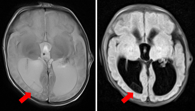
Observations: The authors present a case of intraventricular migration of Gelfoam after ventriculoperitoneal shunt placement in a 6-week-old infant. The infant was imaged regularly after ventriculoperitoneal shunt placement, and the Gelfoam sponge persisted within the ventricles on all images until 11 months after surgery. At no time during follow-up did the patient have any symptoms of hydrocephalus requiring retrieval of the sponge or shunt revision.
Lessons: This is the first case describing time until absorption of a gelatin sponge within the ventricle and successful conservative management.
Keywords: Gelfoam; hydrocephalus; neurosurgery; shunt.
Conflict of interest statement
Figures


Similar articles
-
Gelfoam Migration: A Potential Cause of Recurrent Hydrocephalus.World Neurosurg. 2020 Oct;142:212-217. doi: 10.1016/j.wneu.2020.06.214. Epub 2020 Jul 4. World Neurosurg. 2020. PMID: 32634637
-
Comparing fourth ventricle shunt survival after placement via stereotactic transtentorial and suboccipital approaches.J Neurosurg Pediatr. 2013 Jun;11(6):623-9. doi: 10.3171/2013.3.PEDS12442. Epub 2013 Apr 19. J Neurosurg Pediatr. 2013. PMID: 23601013
-
Revision rate of pediatric ventriculoperitoneal shunts after 15 years.J Neurosurg Pediatr. 2013 Jan;11(1):15-9. doi: 10.3171/2012.9.PEDS1298. Epub 2012 Oct 26. J Neurosurg Pediatr. 2013. PMID: 23101557
-
Rate and Risk Factors for Shunt Revision in Pediatric Patients with Hydrocephalus-A Population-Based Study.World Neurosurg. 2017 May;101:615-622. doi: 10.1016/j.wneu.2017.02.030. Epub 2017 Feb 14. World Neurosurg. 2017. PMID: 28213196 Review.
-
Fourth ventricular entrapment caused by rostrocaudal herniation following shunt malfunction.Pediatr Neurosurg. 1993 Jul-Aug;19(4):209-14. doi: 10.1159/000120733. Pediatr Neurosurg. 1993. PMID: 8329307 Review.
References
-
- Jenkins HP, Janda R, Clarke J. Clinical and experimental observations on the use of gelatin sponge or foam. Surgery. 1946;20(1):124–132. - PubMed
-
- Jenkins HP, Senz EH, Owen HW, Jampolis RW. Present status of gelatin sponge for the control of hemorrhage; with experimental data on its use for wounds of the great vessels and the heart. J Am Med Assoc. 1946;132(11):614–619. - PubMed
-
- Treves N. Prophylaxis of postmammectomy lymphedema by the use of gelfoam laminated rolls; a preliminary report, with a review of the theories on the etiology of elephantiasis chirurgica and a summary of previous operations for its control. Cancer. 1952;5(1):73–84. - PubMed
-
- Barnes AC. The use of gelatin foam sponges in obstetrics and gynecology. Am J Obstet Gynecol. 1947;54(1):105–107. - PubMed
-
- Barbolt TA, Odin M, Léger M, Kangas L. Pre-clinical subdural tissue reaction and absorption study of absorbable hemostatic devices. Neurol Res. 2001;23(5):537–542. - PubMed
LinkOut - more resources
Full Text Sources
