

Meningioma with holo-sagittal sinus involvement treated successfully with intrinsic sinus surgery: illustrative case
- PMID: 36303492
- PMCID: PMC9379721
- DOI: 10.3171/CASE21710
Meningioma with holo-sagittal sinus involvement treated successfully with intrinsic sinus surgery: illustrative case
Abstract
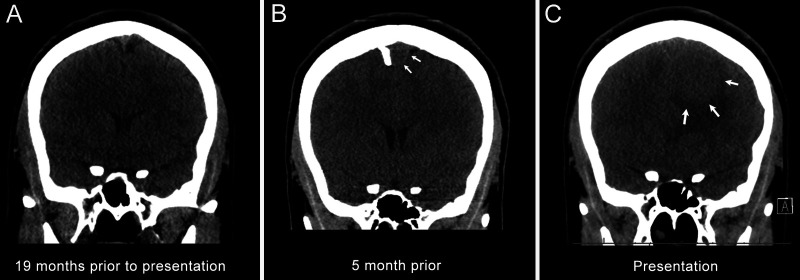
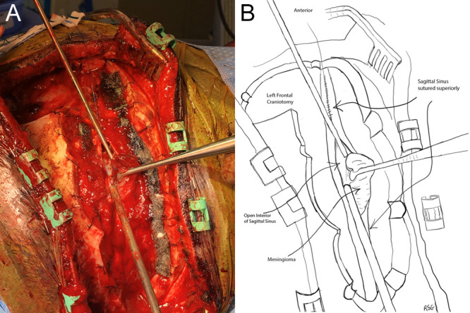
Background: This report describes an unusual meningioma with a large left frontal component and extensive growth within the sagittal sinus and its successful treatment with a staged approach: left frontal craniotomy followed by a sagittal craniotomy and intrinsic removal of the tumor from the sagittal sinus.
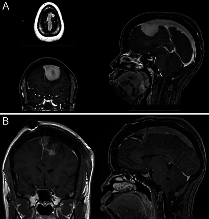
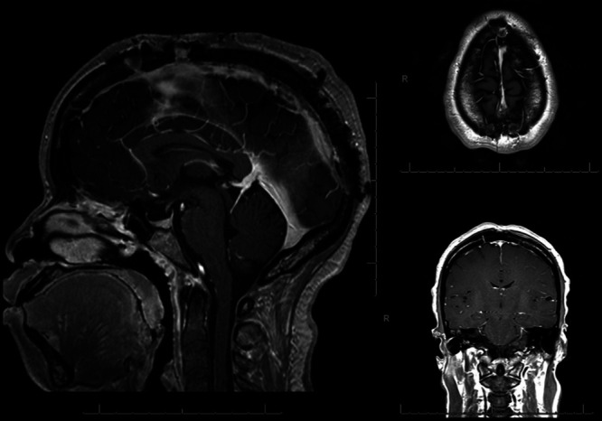
Observations: A previously healthy 27-year-old presented with 6 months of progressively worsening bilateral headaches, visual changes, and nausea. On examination she had a left cranial nerve VI palsy and severe papilledema. Magnetic resonance imaging revealed a 5.1 × 3.8 × 4.1 cm homogenously enhancing left superior frontal parafalcine extra-axial mass with surrounding vasogenic edema and growth through the sagittal sinus extending just short of the torcula.
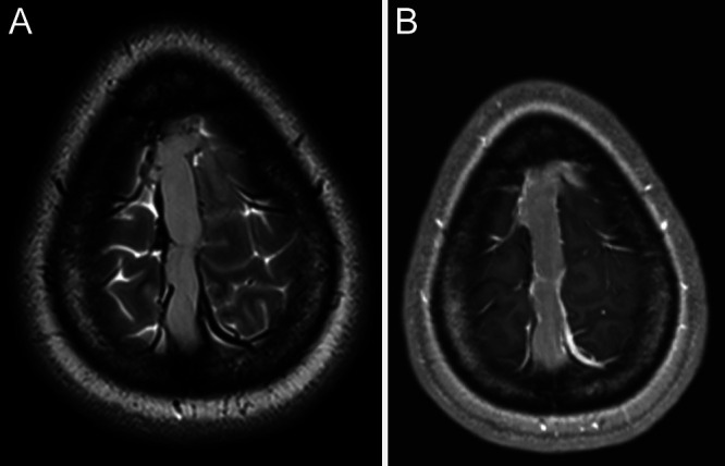
Lessons: This case report describes a fast-growing meningioma with a unique pattern of spread, growing through the sagittal sinus as if it were a conduit and resulting in complete occlusion of flow in the sinus. An important recognition in this case was that a robust parasagittal venous plexus had developed on either side of the falx cerebri with drainage to the inferior sagittal sinus. This collateral drainage pattern allowed for an extradural opening of the sagittal sinus from front to back and intrinsic resection of the tumor from the sinus with preservation of the lateral walls of the sinus.
Keywords: case report; meningioma; sagittal sinus; sinus surgery.
Conflict of interest statement
Figures
Similar articles
-
Challenging Resection of Bilateral Parasagittal and Falcine Meningioma Involving Both Anterior Third and Middle Third of the Superior Sagittal Sinus: A Case Report and Literature Review.Cureus. 2024 Jul 18;16(7):e64865. doi: 10.7759/cureus.64865. eCollection 2024 Jul. Cureus. 2024. PMID: 39156289 Free PMC article.
-
Surgical management of a transosseous meningioma with invasion of torcula, superior sagittal sinus, transverse sinus, calvaria, and scalp.Surg Neurol Int. 2015 Mar 20;6:40. doi: 10.4103/2152-7806.153708. eCollection 2015. Surg Neurol Int. 2015. PMID: 25883832 Free PMC article.
-
Dural arteriovenous fistula associated with superior sagittal sinus occlusion secondary to invasion by a parafalcine meningioma: case report.Neurosurgery. 2010 Jul;67(1):205-7; discussion 207. doi: 10.1227/01.NEU.0000370089.94032.4F. Neurosurgery. 2010. PMID: 20559067
-
Fatal Superior Sagittal Sinus and Torcular Thrombosis After Vestibular Schwannoma Surgery: Report of a Rare Complication and Review of the Literature.World Neurosurg. 2016 Dec;96:607.e19-607.e24. doi: 10.1016/j.wneu.2016.09.075. Epub 2016 Sep 28. World Neurosurg. 2016. PMID: 27686505 Review.
-
Idiopathic Hypertrophic Pachymeningitis Mimicking Meningioma with Occlusion of Superior Sagittal Sinus: Case Report and Review of Literature.World Neurosurg. 2019 Jul;127:534-537. doi: 10.1016/j.wneu.2019.04.006. Epub 2019 Apr 6. World Neurosurg. 2019. PMID: 30965168 Review.
Cited by
-
Challenging Resection of Bilateral Parasagittal and Falcine Meningioma Involving Both Anterior Third and Middle Third of the Superior Sagittal Sinus: A Case Report and Literature Review.Cureus. 2024 Jul 18;16(7):e64865. doi: 10.7759/cureus.64865. eCollection 2024 Jul. Cureus. 2024. PMID: 39156289 Free PMC article.
References
-
- Haddad GF, Al-Mefty O, Aboulrauf SI. Youmans Neurological Surgery. 5th ed. Saunders; 2004. pp. 1099–1131.
-
- Sindou MP, Alvernia JE. Results of attempted radical tumor removal and venous repair in 100 consecutive meningiomas involving the major dural sinuses. J Neurosurg. 2006;105(4):514–525. - PubMed
-
- Sindou M. Meningiomas invading the sagittal or transverse sinuses, resection with venous reconstruction. J Clin Neurosci. 2001;8(1) suppl 1:8–11. - PubMed
LinkOut - more resources
Full Text Sources
