

The relevance of sperm morphology in male infertility
- PMID: 36303645
- PMCID: PMC9580829
- DOI: 10.3389/frph.2022.945351
The relevance of sperm morphology in male infertility
Abstract
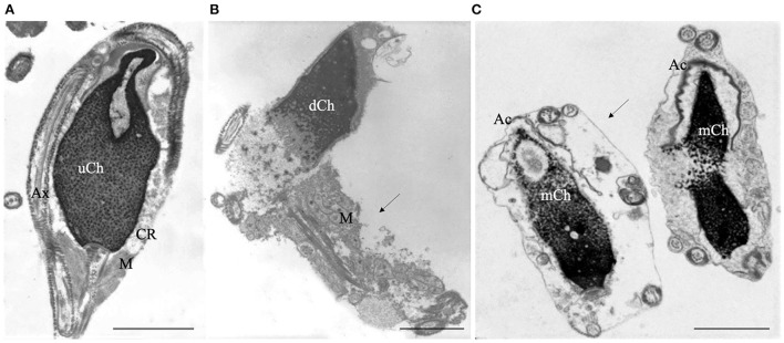
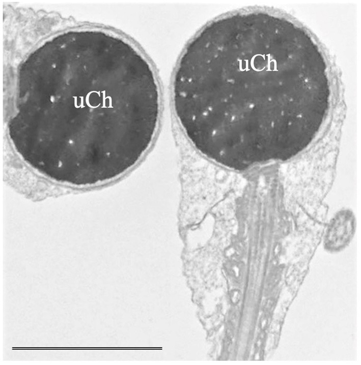
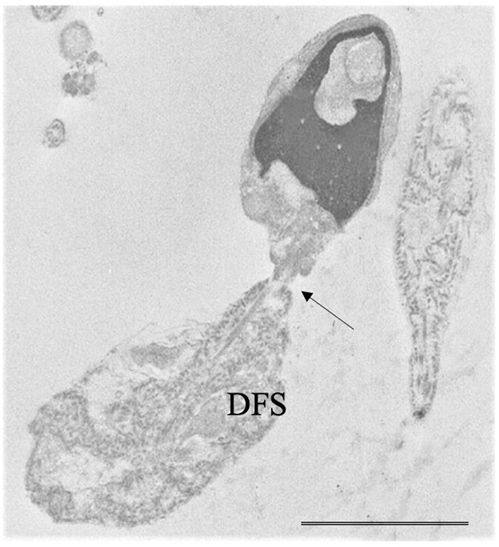
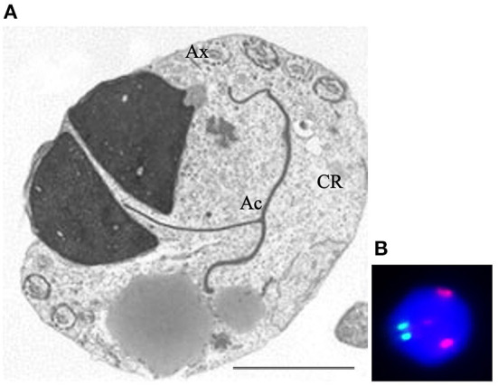
This brief report concerns the role of human sperm morphology assessment in different fields of male infertility: basic research, genetics, assisted reproduction technologies, oxidative stress. One of the best methods in studying sperm morphology is transmission electron microscopy (TEM) that enables defining the concept of sperm pathology and classifying alterations in non-systematic and systematic. Non-systematic sperm defects affect head and tail in variable ratio, whereas the rare systematic defects are characterized by a particular anomaly that marks most sperm of an ejaculate. TEM analysis and fluorescence in situ hybridization represent outstanding methods in the study of sperm morphology and cytogenetic in patients with altered karyotype characterizing their semen quality before intracytoplasmic sperm injection. In recent years, the genetic investigations on systematic sperm defects, made extraordinary progress identifying candidate genes whose mutations induce morphological sperm anomalies. The question if sperm morphology has an impact on assisted fertilization outcome is debated. Nowadays, oxidative stress represents one of the most important causes of altered sperm morphology and function and can be analyzed from two points of view: 1) spermatozoa with cytoplasmic residue produce reactive oxygen species, 2) the pathologies with inflammatory/oxidative stress background cause morphological alterations. Finally, sperm morphology is also considered an important endpoint in in vitro experiments where toxic substances, drugs, antioxidants are tested. We think that the field of sperm morphology is far from being exhausted and needs other research. This parameter can be still considered a valuable indicator of sperm dysfunction both in basic and clinical research.
Keywords: assisted reproduction technologies (ART); genetics; human sperm; oxidative stress; sperm morphology; systematic sperm defects; transmission electron microscopy.
Copyright © 2022 Moretti, Signorini, Noto, Corsaro and Collodel.
Conflict of interest statement
The authors declare that the research was conducted in the absence of any commercial or financial relationships that could be construed as a potential conflict of interest.
Figures
References
-
- World Health Organization (WHO) . WHO Manual for the Laboratory Examination and Processing of Human Semen. 6th ed. WHO press: Geneva Switzerland; (2021).
-
- Agarwal A, Sharma R, Gupta S, Finelli R, Parekh N, Panner Selvam MK, et al. Sperm morphology assessment in the era of intracytoplasmic sperm injection: reliable results require focus on standardization, quality control, and training. World J Mens Health. (2021) 40:347–60. 10.5534/wjmh.210054 - DOI - PMC - PubMed
-
- World Health Organization (WHO) . WHO Laboratory Manual for the Examination of Human Semen and Sperm-Cervical Mucus Interaction. 4th ed. Cambridge: Cambridge University Press; (1999).
LinkOut - more resources
Full Text Sources