Review
. 2022 May 4:4:863173.
doi: 10.3389/frph.2022.863173. eCollection 2022.

Non-Invasive and Mechanism-Based Molecular Assessment of Endometrial Receptivity During the Window of Implantation: Current Concepts and Future Prospective Testing Directions

Affiliations
Review

Non-Invasive and Mechanism-Based Molecular Assessment of Endometrial Receptivity During the Window of Implantation: Current Concepts and Future Prospective Testing Directions

Bei Sun et al. Front Reprod Health. .

Abstract

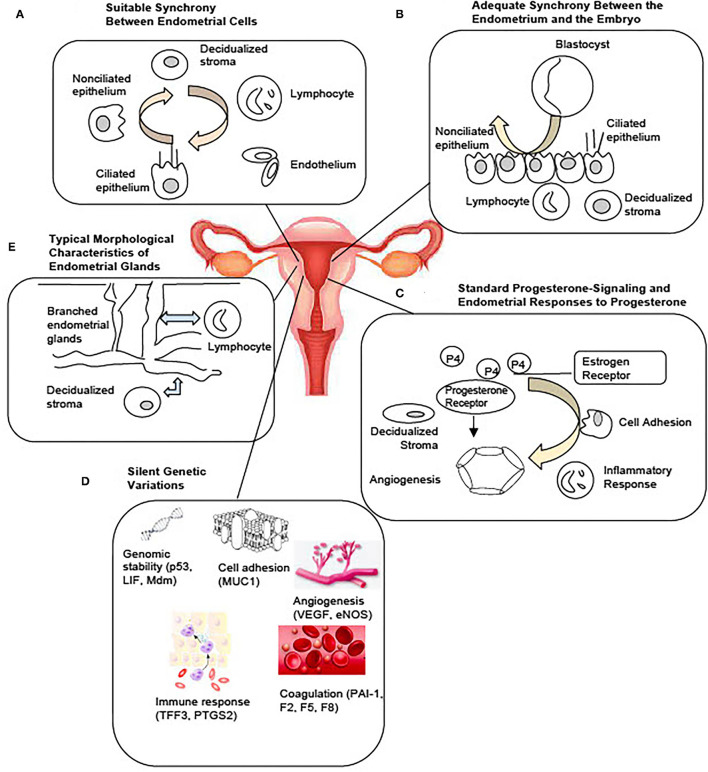
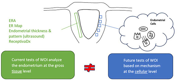
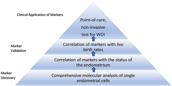
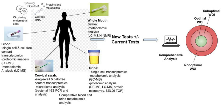
Suboptimal endometrial receptivity and altered embryo-endometrial crosstalk account for approximately two-thirds of human implantation failures. Current tests of the window of implantation, such as endometrial thickness measurements and the endometrial receptivity assay, do not consistently improve clinical outcomes as measured by live birth rates. Understanding the mechanisms regulating the endometrial receptivity during the window of implantation is a critical step toward developing clinically meaningful tests. In this narrative review, the available literature is evaluated regarding mechanisms that regulate the endometrial receptivity during the window of implantation and the current tests developed. Overall, both animal and human studies point to five possible and interrelated mechanisms regulating the endometrial window of implantation: suitable synchrony between endometrial cells, adequate synchrony between the endometrium and the embryo, standard progesterone signaling and endometrial responses to progesterone, silent genetic variations, and typical morphological characteristics of the endometrial glands. The biological basis of current clinical markers or tests of window of implantation is poor. Future studies to elucidate the mechanisms shaping the window of implantation and to investigate the potential markers based on these mechanisms are required. In addition, molecular testing of the endometrium at single-cell resolution should be an initial step toward developing clinically meaningful tests for the optimal window of implantation. As understanding of the optimal window of implantation continues to evolve, one can envision the future development of non-invasive, mechanism-based testing of the window of implantation.

Keywords: endometrial receptivity; mechanism; molecular markers; non-invasive testing; point-of-care testing; window of implantation (WOI).


Conflict of interest statement

The authors declare that the research was conducted in the absence of any commercial or financial relationships that could be construed as a potential conflict of interest.

Figures

Figure 1
Figure 1
Potential Mechanisms Regulating the Optimal Window of Implantation. Based on current literature, five possible and interrelated mechanisms include: (A) Suitable synchrony between the endometrial cells. (B) Adequate synchrony between the endometrium and the embryo. (C) Standard progesterone-signaling and the endometrial responses to progesterone. (D) Silent genetic variations. (E) Typical morphological characteristics of the endometrial glands may together constitute the molecular basis of a receptive endometrium during the window of implantation.
Figure 2
Figure 2
Current tests of the window of implantation are not based on full knowledge of the mechanisms regulating the endometrial receptivity. Activities of the endometrial cells underly the processes essential to the entry and maintenance of the window of implantation. Commercial tests of window of implantation, however, do not analyze the endometrium at the cellular level necessary to investigate the status of receptivity.
Figure 3
Figure 3
Roadmap to develop proposed tests for the window of implantation. The first stage of the roadmap involves marker discovery through comprehensive single-cell molecular analysis of the endometrial cells. Markers specific to each endometrial cell type associated with histological findings of receptivity and live birth can be validated and thresholds of the markers can be established through large-scale clinical studies. The second stage involves development of point-of-care, non-invasive tests for the window of implantation. This stage requires selection of essential markers from different sources such as blood and urine tests to be integrated into routine clinical use.
Figure 4
Figure 4
Development of non-invasive tests for the optimal window of implantation (WOI). Multiple analyses could be performed including transcriptomic, metabolomic, proteomic and microbiome profiling through routine saliva, urine, blood or cervical swab tests. The proposed tests above are all currently technically feasible and could be developed into commercial tests. These tests offer the opportunity for advancing the understanding of the biological basis of the endometrial receptivity during the WOI. They also encourage future biomarker discovery, validation and integration of the biomarkers of WOI into daily clinical practice for testing of women of reproductive age to improve pregnancy outcomes.

Similar articles

Cited by

References

    1. Duffy JMN, Adamson GD, Benson E, Bhattacharya S, Bofill M, Brian K, et al. . Top 10 priorities for future infertility research: an international consensus development study. Hum Reprod. (2020) 35:2715–24. 10.1093/humrep/deaa242 - DOI - PMC - PubMed
    1. Martel D, Frydman R, Glissant M, Maggioni C, Roche D, Psychoyos A. Scanning electron microscopy of postovulatory human endometrium in spontaneous cycles and cycles stimulated by hormone treatment. J Endocrinol. (1987) 114:319–24. 10.1677/joe.0.1140319 - DOI - PubMed
    1. Næslund G, Lundkvist Ö, Nilsson BO. Transmission electron microscopy of mouse blastocysts activated and growth-arrested in vivo and in vitro. Anat Embryol. (1980) 159:33–48. 10.1007/BF00299253 - DOI - PubMed
    1. Psychoyos A. Uterine receptivity for nidation. Ann N Y Acad Sci. (1986) 476:36–42. 10.1111/j.1749-6632.1986.tb20920.x - DOI - PubMed
    1. Paria BC, Huet-Hudson YM, Dey SK. Blastocyst's state of activity determines the “window” of implantation in the receptive mouse uterus. Proc Natl Acad Sci U S A. (1993) 90:10159–62. 10.1073/pnas.90.21.10159 - DOI - PMC - PubMed