

Association of microRNAs With Embryo Development and Fertilization in Women Undergoing Subfertility Treatments: A Pilot Study
- PMID: 36303988
- PMCID: PMC9580729
- DOI: 10.3389/frph.2021.719326
Association of microRNAs With Embryo Development and Fertilization in Women Undergoing Subfertility Treatments: A Pilot Study
Abstract
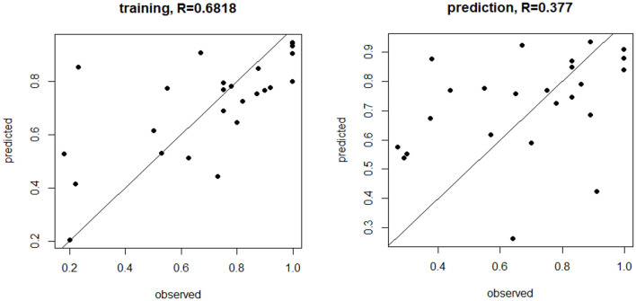
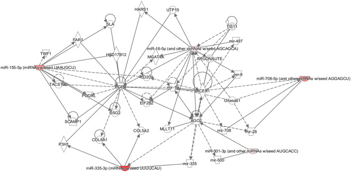
Objective: Small non-coding RNAs, known as microRNAs (miRNAs), have emerging regulatory functions within the ovary that have been related to fertility. This study was undertaken to determine if circulating miRNAs reflect the changes associated with the parameters of embryo development and fertilization. Methods: In this cross-sectional pilot study. Plasma miRNAs were collected from 48 sequentially presenting women in the follicular phase prior to commencing in vitro fertilization (IVF). Circulating miRNAs were measured using locked nucleic acid (LNA)-based quantitative PCR (qPCR), while an updated miRNA data set was used to determine their level of expression. Results: Body mass index and weight were associated with the miRNAs let7b-3p and miR-375, respectively (p < 0.05), with the same relationship being found between endometrium thickness at oocyte retrieval and miR-885-5p and miR-34a-5p (p < 0.05). In contrast, miR-1260a was found to be inversely associated with anti-Mullerian hormone (AMH; p = 0.007), while miR-365a-3p, miR122-5p, and miR-34a-5p correlated with embryo fertilization rates (p < 0.05). However, when omitting cases of male infertility (n = 15), miR122-5p remained significant (p < 0.05), while miR-365a-3p and miR-34a-5p no longer differed; interestingly, however, miR1260a and mir93.3p became significant (p = 0.0087/0.02, respectively). Furthermore, age was negatively associated with miR-335-3p, miR-28-5p, miR-155-5p, miR-501-3p, and miR-497-5p (p < 0.05). Live birth rate was negatively associated with miR-335-3p, miR-100-5p, miR-497-5p, let-7d, and miR-574-3p (p < 0.05), but these were not significant when age was accounted for.However, with the exclusion of male factor infertility, all those miRNAs were no longer significant, though miR.150.5p emerged as significant (p = 0.042). A beta-regression model identified miR-1260a, miR-486-5p, and miR-132-3p (p < 0.03, p = 0.0003, p < 0.00001, respectively) as the most predictive for fertilization rate. Notably, changes in detectable miRNAs were not linked to cleavage rate, top quality embryos (G3D3), and blastocyst or antral follicle count. An ingenuity pathway analysis showed that miRNAs associated with age were also associated with the variables found in reproductive system diseases. Conclusion: Plasma miRNAs prior to the IVF cycle were associated with differing demographic and IVF parameters, including age, and may be predictive biomarkers of fertilization rate.
Keywords: IVF; embryo; fertilization rate; infertility; microRNA.
Copyright © 2021 Butler, Cunningham, Ramachandran, Diboun, Halama, Sathyapalan, Najafi-Shoushtari and Atkin.
Conflict of interest statement
The authors declare that the research was conducted in the absence of any commercial or financial relationships that could be construed as a potential conflict of interest.
Figures
Similar articles
-
Expression of microRNA in follicular fluid in women with and without PCOS.Sci Rep. 2019 Nov 8;9(1):16306. doi: 10.1038/s41598-019-52856-5. Sci Rep. 2019. PMID: 31705013 Free PMC article.
-
Roles of miR-10a-5p and miR-103a-3p, Regulators of BDNF Expression in Follicular Fluid, in the Outcomes of IVF-ET.Front Endocrinol (Lausanne). 2021 May 12;12:637384. doi: 10.3389/fendo.2021.637384. eCollection 2021. Front Endocrinol (Lausanne). 2021. PMID: 34054723 Free PMC article.
-
hsa-miR-320a-3p and hsa-miR-483-5p levels in human granulosa cells: promising bio-markers of live birth after IVF/ICSI.Reprod Biol Endocrinol. 2022 Nov 21;20(1):160. doi: 10.1186/s12958-022-01037-7. Reprod Biol Endocrinol. 2022. PMID: 36411450 Free PMC article.
-
Identifying the differentially expressed peripheral blood microRNAs in psychiatric disorders: a systematic review and meta-analysis.Front Psychiatry. 2024 May 17;15:1390366. doi: 10.3389/fpsyt.2024.1390366. eCollection 2024. Front Psychiatry. 2024. PMID: 38827444 Free PMC article.
-
Investigating the Imperative Role of microRNAs Expression in Human Embryo Implantation: A Narrative Review Based on Recent Evidence.Biomedicines. 2024 Nov 15;12(11):2618. doi: 10.3390/biomedicines12112618. Biomedicines. 2024. PMID: 39595182 Free PMC article. Review.
Cited by
-
Circulating miRNAs in the first trimester and pregnancy complications: a systematic review.Epigenetics. 2023 Dec;18(1):2152615. doi: 10.1080/15592294.2022.2152615. Epub 2022 Dec 12. Epigenetics. 2023. PMID: 36503407 Free PMC article.
-
Identification of Differentially Expressed mRNAs and miRNAs and Related Regulatory Networks in Cumulus Oophorus Complexes Associated with Fertilization.Reprod Sci. 2024 May;31(5):1408-1419. doi: 10.1007/s43032-023-01413-7. Epub 2024 Jan 12. Reprod Sci. 2024. PMID: 38216777
-
miRNA expression profiles of peripheral white blood cells from beef heifers with varying reproductive potential.Front Genet. 2023 May 10;14:1174145. doi: 10.3389/fgene.2023.1174145. eCollection 2023. Front Genet. 2023. PMID: 37234872 Free PMC article.
-
Bone marrow mesenchymal stem cell-derived exosomes shuttle microRNAs to endometrial stromal fibroblasts that promote tissue proliferation /regeneration/ and inhibit differentiation.Stem Cell Res Ther. 2024 May 1;15(1):129. doi: 10.1186/s13287-024-03716-1. Stem Cell Res Ther. 2024. PMID: 38693588 Free PMC article.
-
Meta-analysis of endometrial transcriptome data reveals novel molecular targets for recurrent implantation failure.J Assist Reprod Genet. 2024 May;41(5):1417-1431. doi: 10.1007/s10815-024-03077-x. Epub 2024 Mar 8. J Assist Reprod Genet. 2024. PMID: 38456991 Free PMC article.
References
Associated data
LinkOut - more resources
Full Text Sources
Other Literature Sources