

Case report: Characterization of a rare pathogenic variant associated with loss of COL3A1 expression in vascular Ehlers Danlos syndrome
- PMID: 36304539
- PMCID: PMC9595653
- DOI: 10.3389/fcvm.2022.939013
Case report: Characterization of a rare pathogenic variant associated with loss of COL3A1 expression in vascular Ehlers Danlos syndrome
Abstract
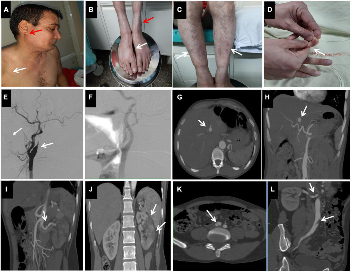
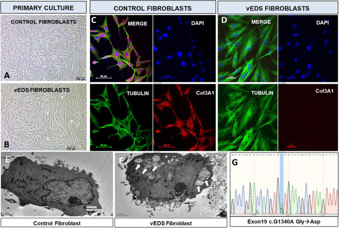
The vascular subtype of Ehlers Danlos Syndrome (vEDS) is a rare connective tissue disorder characterized by spontaneous arterial, bowel or organ rupture. The diagnosis of vEDS is established in a proband by identification of a heterozygous pathogenic variant in the alpha-1 gene of type III collagen (COL3A1) by molecular analysis. In this report, we present a case of vEDS with life threatening, spontaneous arterial dissections in association with an uncharacterized rare variant of COL3A1, exon19:c.1340G > A. Primary culture of patient skin fibroblasts followed by immunofluorescence revealed a complete absence of COL3A1 protein expression as well as altered morphology. Electron microscopy of the cultured fibroblasts showed abnormal vacuoles in the cytoplasm suggestive of a secretory defect. In this study, we have performed functional characterization of the COL3A1 exon19:c.1340G > A variant for the first time and this may now be classified as likely pathogenic in vEDS. *Both JM and LRL contributed equally in the manuscript and should both be considered as the first author.
Keywords: COL3A1 pathogenic variant; clinical fibroblast testing; exome sequencing; hepatic artery dissection; stroke; vascular Ehlers Danlos syndrome.
Copyright © 2022 Manhas, Lohani, Seethy, Kumar, Gamanagatti and Sen.
Conflict of interest statement
The authors declare that the research was conducted in the absence of any commercial or financial relationships that could be construed as a potential conflict of interest.
Figures
Similar articles
-
A multi-institutional experience in vascular Ehlers-Danlos syndrome diagnosis.J Vasc Surg. 2020 Jan;71(1):149-157. doi: 10.1016/j.jvs.2019.04.487. Epub 2019 Jul 26. J Vasc Surg. 2020. PMID: 31353273 Free PMC article.
-
Splenic artery pathology presentation, operative interventions, and outcomes in 88 patients with vascular Ehlers-Danlos syndrome.J Vasc Surg. 2023 Aug;78(2):394-404. doi: 10.1016/j.jvs.2023.04.007. Epub 2023 Apr 15. J Vasc Surg. 2023. PMID: 37068529
-
A multi-institutional experience in the aortic and arterial pathology in individuals with genetically confirmed vascular Ehlers-Danlos syndrome.J Vasc Surg. 2019 Nov;70(5):1543-1554. doi: 10.1016/j.jvs.2019.01.069. Epub 2019 May 21. J Vasc Surg. 2019. PMID: 31126764 Free PMC article.
-
Vascular Ehlers-Danlos Syndrome With a Novel Missense COL3A1 Mutation Present With Pulmonary Complications and Iliac Arterial Dissection.Vasc Endovascular Surg. 2018 Feb;52(2):138-142. doi: 10.1177/1538574417745432. Epub 2017 Dec 7. Vasc Endovascular Surg. 2018. PMID: 29216800 Review.
-
A Novel Frameshift COL3A1 Variant in Vascular Ehlers-Danlos Syndrome.Ann Vasc Surg. 2019 Nov;61:472.e9-472.e13. doi: 10.1016/j.avsg.2019.05.057. Epub 2019 Aug 5. Ann Vasc Surg. 2019. PMID: 31394236 Review.
Cited by
-
Characterizing the immune response to myocardial infarction in pigs.Basic Res Cardiol. 2024 Jun;119(3):453-479. doi: 10.1007/s00395-024-01036-2. Epub 2024 Mar 15. Basic Res Cardiol. 2024. PMID: 38491291 Free PMC article.
-
Spontaneous celiac artery aneurysms in 13-year-old and 10-year-old brothers with PLOD1-related kyphoscoliotic Ehlers-Danlos syndrome.J Vasc Surg Cases Innov Tech. 2024 Mar 21;10(3):101465. doi: 10.1016/j.jvscit.2024.101465. eCollection 2024 Jun. J Vasc Surg Cases Innov Tech. 2024. PMID: 38694482 Free PMC article.
References
-
- Malfait F, Francomano C, Byers P, Belmont J, Berglund B, Black J, et al. The 2017 international classification of the ehlers-danlos syndromes. Am J Med Genet C Semin Med Genet. (2017) 175:8–26. - PubMed
-
- Byers PH. Vascular ehlers-danlos syndrome. In: Adam MP, Ardinger HH, Pagon RA, Wallace SE, Bean LJ, Stephens K. editors. GeneReviews. Seattle (WA): University of Washington; (2019). - PubMed
-
- The Ehlers–Danlos Syndromes, Rare Types - Brady - 2017. American Journal of Medical Genetics Part C: Seminars in Medical Genetics - Wiley Online Library. (2020). Available online at: https://onlinelibrary.wiley.com/doi/full/10.1002/ajmg.c.31550 (accessed July 24, 2020). - DOI - PubMed
Publication types
LinkOut - more resources
Full Text Sources
Miscellaneous