

Case Reports
doi: 10.1080/08998280.2022.2104528.
eCollection 2022.
Diagnosing aortic syphilis
Affiliations
- PMID: 36304622
- PMCID: PMC9586584
- DOI: 10.1080/08998280.2022.2104528
Item in Clipboard
Case Reports
Diagnosing aortic syphilis
Proc (Bayl Univ Med Cent).
.
Abstract
Described herein is a morbidly obese 57-year-old man with an aneurysm involving the tubular portion of the aorta. Examination of the wall of the operatively resected aneurysm disclosed classic findings of aortic syphilis, a condition that clearly has not disappeared. If there is an aneurysm involving the tubular portion of the ascending aorta in the absence of aortic dissection or involvement of the sinuses of Valsalva, the most likely diagnosis is aortic syphilis. In these circumstances, the serologic test for syphilis is often negative.
Keywords: Aortic aneurysm; aortic operation; aortic syphilis.
Copyright © 2022 Baylor University Medical Center.
Figures

(a) Diagram of the aortic aneurysm before and after operative therapy. (b) View of the dilated ascending aorta before and after its replacement.

View of the aortogram showing the (a, b) dilated tubular portion of the aorta. The sinus of Valsalva (SV) is of normal size. AA indicates ascending aorta; DTA, descending thoracic aorta. (c) Opened aorta showing the extensive fibrous and calcific deposits on the intimal surface. The circumference of the aorta is 16 cm and, when intact, the diameter is 5.1 cm. (d) Photomicrograph of the wall of the fusiform aneurysm showing large collections of cholesterol clefts in the intima (I) and destroyed part of the media (M). The adventitia (A) is thickened by fibrous tissue within which are collections of lymphocytes and plasmacytes (small arrows). Movat stain, ×20.
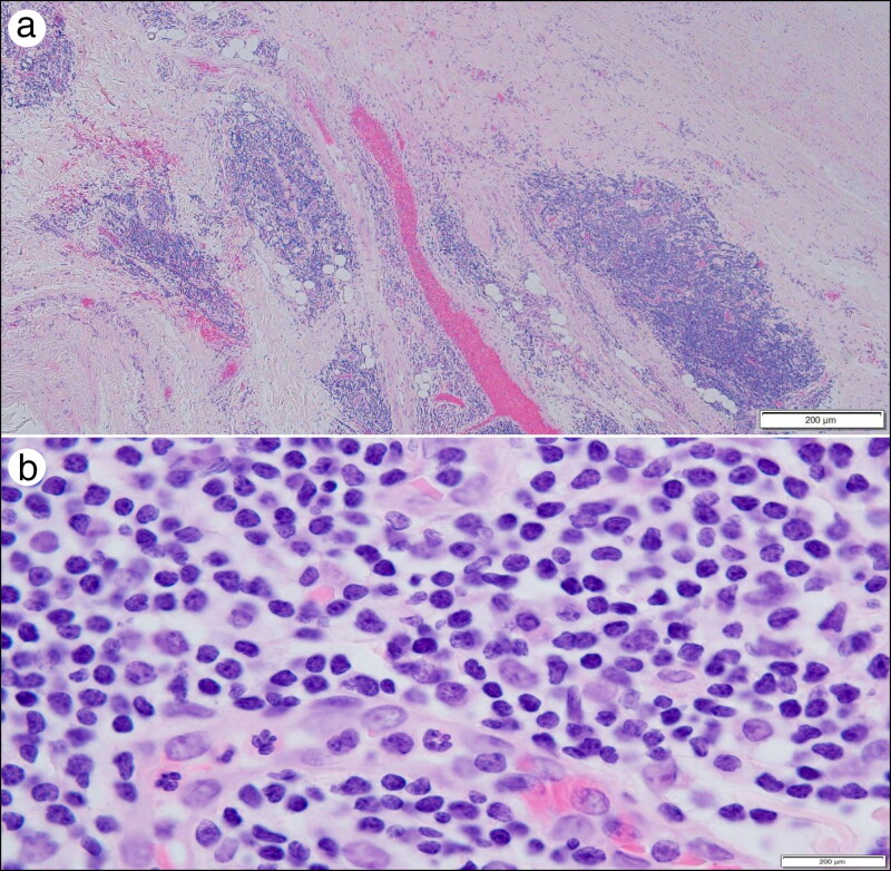
Photomicrographs of portions of the aortic media showing (a) large clumps of lymphocytes and plasmacytes and (b) a close-up of cells in the clumps. Hematoxylin-eosin stains, × 20 (a) and ×1000 (b).
References
Publication types
LinkOut - more resources
Full Text Sources